









Mitochondrial Metabolism in Neural Tissue: The Impact of Methylene Blue on the Respiratory Chain and ATP Production
Part 1: Foundations of Neural Bioenergetics and the Redox Chemistry of Methylene Blue
Neural tissue is metabolically unusual. Neurons must continuously preserve steep transmembrane ion gradients while supporting action potentials, synaptic transmission, neurotransmitter recycling, calcium regulation, axonal transport and structural maintenance. These processes require a continuous supply of adenosine triphosphate, or ATP.
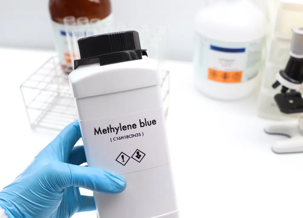
Methylene blue is relevant to neural bioenergetics because it is a redox-active phenothiazinium compound capable of accepting and donating electrons. In experimental systems, this chemistry allows methylene blue to interact with NADH-dependent reactions, cytochrome c, molecular oxygen and components of the mitochondrial respiratory chain. However, the biological consequences are highly dependent on concentration, redox environment, oxygen availability, mitochondrial condition and experimental model.
This first part establishes the biochemical framework required to interpret later claims about methylene blue. It does not assume that increased oxygen consumption automatically means increased ATP production, nor that an effect demonstrated in isolated mitochondria must occur identically in intact human neural tissue.
Scope and Scientific Evidence Framework
Claims concerning methylene blue should be separated according to the model in which they were demonstrated.
| Evidence category | What is directly measured | What cannot automatically be concluded |
|---|---|---|
| Chemical system | Electron transfer, reduction potential, spectral change | Cellular or clinical benefit |
| Purified enzyme system | Interaction with a particular enzyme or cofactor | Whole-mitochondrial function |
| Isolated mitochondria | Respiration, membrane potential, ATP synthesis, ROS | Effects in intact neurons or humans |
| Cultured neural cells | Viability, respiration, ATP, signalling | Organ-level pharmacology |
| Brain slices or synaptosomes | Local neural metabolism and synaptic effects | Whole-organism outcomes |
| Animal model | Tissue, behavioural and physiological effects | Human efficacy |
| Human metabolic study | Cerebral blood flow, oxygen use or metabolite changes | Long-term clinical benefit |
| Controlled clinical trial | Defined clinical outcomes | Universal effects outside the studied population |
An observation should therefore be described as, for example, “demonstrated in isolated guinea-pig brain mitochondria” rather than simply “methylene blue increases brain ATP.”
Why Neural Tissue Has Exceptional Energy Requirements
Neurons operate far from electrochemical equilibrium. Intracellular sodium is kept low relative to extracellular sodium, while intracellular potassium is kept high. Calcium concentrations are controlled across an even steeper gradient.
These gradients are essential for electrical signalling, but they are continuously disturbed by ion-channel activity. Every action potential allows sodium and potassium ions to move down their electrochemical gradients. Synaptic receptor activation adds further sodium, calcium and other ionic currents. Restoring the original gradients requires ATP-dependent transport.
An influential quantitative energy budget for rodent grey matter estimated that action potentials and postsynaptic glutamatergic signalling represented major components of signalling-related energy expenditure. The exact percentages depend on assumptions, neuronal type and firing pattern, but the central conclusion remains that information processing is energetically expensive.
Major ATP-Dependent Processes in Neurons
| Process | Principal ATP-dependent mechanism | Consequence of ATP failure |
|---|---|---|
| Restoration of Na⁺ and K⁺ gradients | Na⁺/K⁺-ATPase | Membrane depolarisation and impaired firing |
| Calcium extrusion | Plasma-membrane Ca²⁺-ATPase and indirect Na⁺/Ca²⁺ exchange | Cytosolic calcium accumulation |
| Endoplasmic-reticulum calcium storage | SERCA pumps | Disturbed intracellular calcium signalling |
| Synaptic-vesicle acidification | Vacuolar H⁺-ATPase | Impaired neurotransmitter loading |
| Vesicle recycling | ATP- and GTP-dependent trafficking | Reduced synaptic transmission |
| Axonal transport | Kinesin- and dynein-associated ATP hydrolysis | Failure of organelle and cargo delivery |
| Protein quality control | Chaperones and proteasomal systems | Misfolded-protein accumulation |
| Lipid and membrane maintenance | Biosynthetic and remodelling reactions | Loss of membrane integrity |
| Neurotransmitter recycling | Transport and enzymatic conversion | Disrupted synaptic homeostasis |
| Mitochondrial dynamics | Fusion, fission and mitophagy machinery | Abnormal mitochondrial distribution |
The Na⁺/K⁺-ATPase as a Central Energy Consumer
The Na⁺/K⁺-ATPase hydrolyses one ATP molecule during a transport cycle that exports three sodium ions and imports two potassium ions. Because the transported charges are unequal, the pump is electrogenic.
Its function is not merely to establish the resting membrane potential. It also restores gradients after:
- action potentials;
- excitatory postsynaptic currents;
- sodium-dependent neurotransmitter transport;
- secondary active transport;
- astrocytic clearance of extracellular potassium;
- astrocytic uptake of glutamate.
The greater the net ionic movement during signalling, the greater the ATP requirement for restoring homeostasis.
Simplified Pump Reaction
Experimental Puzzle 1
Question: A neuron continues firing normally for several seconds after oxidative phosphorylation is inhibited. Does this prove that mitochondrial ATP is unnecessary for electrical activity?
Solution: No. The neuron initially retains ATP, phosphocreatine and electrochemical gradients accumulated before inhibition. Glycolysis may also continue temporarily. Electrical failure is delayed until ATP supply becomes insufficient to support ion pumps and other essential processes.
Why Synaptic Transmission Is Energetically Expensive
A chemical synapse requires energy before, during and after neurotransmitter release.
Presynaptically, ATP is needed for:
- vesicle mobilisation;
- vesicle acidification;
- neurotransmitter loading;
- endocytosis and membrane recycling;
- calcium extrusion;
- restoration of sodium gradients;
- local mitochondrial maintenance.
Postsynaptically, receptor-mediated ion currents must be reversed by ion pumps and exchangers. Excitatory glutamatergic transmission can therefore impose a substantial metabolic cost on both the neuron and surrounding astrocytes.
Energy Costs Across a Glutamatergic Synapse
| Compartment | Energy-consuming event | Dominant requirement |
|---|---|---|
| Presynaptic terminal | Action-potential propagation | Restoration of Na⁺ and K⁺ |
| Presynaptic terminal | Ca²⁺ entry | Calcium removal |
| Synaptic vesicle | Proton-gradient formation | ATP hydrolysis by V-type ATPase |
| Presynaptic membrane | Vesicle recycling | ATP/GTP-dependent trafficking |
| Postsynaptic membrane | Ionotropic receptor current | Restoration of ionic gradients |
| Astrocyte | Glutamate uptake | Sodium-gradient restoration |
| Astrocyte | Glutamine synthesis | ATP-dependent amidation |
| Neuron | Glutamine reconversion and transmitter packaging | Enzymatic and transport energy |
Neurons, Astrocytes and Oligodendrocytes Are Metabolically Distinct
“Brain metabolism” is not a single uniform process. Different neural cell types possess different metabolic programmes and responsibilities.
Comparison of Major Neural Cell Types
| Feature | Neurons | Astrocytes | Oligodendrocytes |
|---|---|---|---|
| Primary physiological role | Electrical signalling | Homeostasis and metabolic support | Myelination and axonal support |
| Dependence on oxidative metabolism | Generally high | Flexible; glycolytic and oxidative | Variable by developmental state |
| Glycogen storage | Minimal | Principal neural glycogen store | Limited |
| Direct glucose utilisation | Significant | Significant | Significant |
| Lactate production | Possible | Frequently substantial | Possible |
| Lactate utilisation | Strong oxidative capacity | Can produce or consume lactate | Can support axons metabolically |
| Calcium signalling | Electrical and intracellular | Predominantly intracellular signalling | Regulatory and developmental |
| Vulnerability to ATP failure | Very high | High but often more metabolically flexible | High, particularly for myelin maintenance |
This division is functional rather than absolute. Neurons can use glucose directly, astrocytes contain mitochondria and perform oxidative phosphorylation, and lactate can move in more than one direction depending on concentration gradients and metabolic state.
Cellular Sources of ATP in Neural Tissue
ATP can be generated by two principal mechanisms:
- Substrate-level phosphorylation, especially during glycolysis.
- Oxidative phosphorylation, driven by mitochondrial electron transport.
Glycolysis
Glycolysis converts glucose to pyruvate in the cytosol. Its net products per molecule of glucose are:
- two ATP;
- two NADH;
- two pyruvate.
Glycolysis can generate ATP without direct oxygen consumption. However, sustained glycolytic flux requires regeneration of NAD⁺ from NADH.
When mitochondrial oxidation is limited, pyruvate can be reduced to lactate:
The reaction does not generate additional ATP directly. Its immediate metabolic importance is regeneration of NAD⁺, allowing glycolysis to continue.
Oxidative Metabolism
When pyruvate enters mitochondria, pyruvate dehydrogenase converts it into acetyl-CoA. Acetyl-CoA enters the tricarboxylic-acid cycle, generating:
- NADH;
- FADH₂-linked reducing equivalents;
- GTP or ATP;
- carbon dioxide.
NADH and FAD-linked pathways then provide electrons to the respiratory chain.
Glycolysis Versus Oxidative Phosphorylation
| Property | Glycolysis | Oxidative phosphorylation |
|---|---|---|
| Main location | Cytosol | Inner mitochondrial membrane |
| Direct oxygen requirement | No | Yes, through Complex IV |
| Rate of ATP generation | Potentially rapid | Slower to initiate but high capacity |
| ATP yield per glucose | Low | Much higher |
| NAD⁺ regeneration | Lactate formation or mitochondrial shuttles | Electron transport |
| Primary limitation | Substrate and NAD⁺ availability | Oxygen, substrates and respiratory integrity |
| Major advantage | Can continue during limited mitochondrial function | Efficient ATP production |
| Major risk during failure | Lactate and proton accumulation | Loss of membrane potential and ATP output |
The Astrocyte–Neuron Lactate Shuttle Hypothesis
Pellerin and Magistretti demonstrated that glutamate uptake stimulated glucose utilisation and lactate production in cultured astrocytes. Their findings supported a model in which excitatory neurotransmission activates astrocytic glycolysis, followed by lactate export and neuronal lactate oxidation.
The simplified proposed sequence is:
- A neuron releases glutamate.
- Astrocytic sodium-dependent transporters remove glutamate.
- Sodium entering the astrocyte stimulates Na⁺/K⁺-ATPase activity.
- Energy demand and glycolytic flux increase.
- Astrocytes produce lactate.
- Lactate is exported through monocarboxylate transporters.
- Neurons import and oxidise lactate.
However, the lactate-shuttle model should not be presented as an exclusive or universally unidirectional pathway. Direct activity-dependent neuronal glucose utilisation has also been experimentally observed, and human metabolic studies indicate that lactate exchange can depend on activation state and concentration gradients.
Models of Brain Substrate Distribution
| Model | Astrocytic role | Neuronal role | Scientific limitation |
|---|---|---|---|
| Classical glucose model | Support and homeostasis | Direct glucose oxidation | Understates intercellular lactate exchange |
| Astrocyte-to-neuron lactate shuttle | Glucose uptake and lactate production | Lactate oxidation | May not dominate in every condition |
| Neuron-to-astrocyte lactate transfer | Lactate uptake or disposal | Activity-related lactate production | Direction may vary with metabolic state |
| Bidirectional lactate exchange | Production or consumption | Production or consumption | Hard to resolve spatially in vivo |
| Substrate-flexibility model | Uses glucose, lactate and other fuels | Uses glucose, lactate and ketones | More realistic but less simple |
Experimental Puzzle 2
Question: An experiment detects increased lactate concentration during neuronal activation. Does this prove that astrocytes produced the lactate?
Solution: No. Increased tissue lactate does not identify its cellular source. Lactate could arise from astrocytes, neurons or both. Determining direction requires cell-specific tracers, transporter manipulation, metabolic imaging or isotope-resolved analysis.
Alternative Neural Fuels
Although glucose is the dominant circulating fuel under ordinary conditions, neural tissue can also oxidise:
- lactate;
- ketone bodies;
- pyruvate;
- selected amino-acid-derived carbon skeletons.
Ketone bodies such as β-hydroxybutyrate and acetoacetate can be converted to acetyl-CoA and enter the tricarboxylic-acid cycle. Their importance increases during fasting, prolonged carbohydrate restriction or certain developmental states.
The availability of alternative fuels does not eliminate the requirement for oxygen when ATP is generated through oxidative phosphorylation.
Architecture of the Mitochondrial Respiratory Chain
The respiratory chain is embedded within the inner mitochondrial membrane. It transfers electrons from reduced metabolic cofactors to molecular oxygen while coupling part of the released free energy to proton translocation.
Core Components
| Component | Formal name | Electron donor | Electron acceptor | Direct proton-pumping role |
|---|---|---|---|---|
| Complex I | NADH:ubiquinone oxidoreductase | NADH | Ubiquinone | Yes |
| Complex II | Succinate dehydrogenase | Succinate/FADH₂ pathway | Ubiquinone | No |
| Coenzyme Q | Ubiquinone/ubiquinol pool | Complexes I, II and other enzymes | Complex III | Mobile carrier |
| Complex III | Cytochrome bc₁ complex | Ubiquinol | Cytochrome c | Yes, through Q-cycle chemistry |
| Cytochrome c | Mobile haem protein | Complex III | Complex IV | No direct pumping |
| Complex IV | Cytochrome c oxidase | Reduced cytochrome c | Oxygen | Yes |
| Complex V | F₁F₀-ATP synthase | Proton-motive force | ADP and phosphate | Uses proton flow |
Simplified Electron Pathway
A second entry pathway begins with succinate:
Complex I: NADH:Ubiquinone Oxidoreductase
Complex I oxidises NADH, transfers electrons through flavin mononucleotide and iron–sulphur centres, and reduces ubiquinone to ubiquinol.
The redox reaction is coupled to proton translocation. Experimental work in mammalian systems supports a stoichiometry of approximately four translocated protons for each pair of electrons transferred from NADH.
Functional Consequences of Complex I Activity
- regeneration of mitochondrial NAD⁺;
- entry of NADH-derived electrons into the respiratory chain;
- contribution to the proton-motive force;
- support of tricarboxylic-acid-cycle flux;
- influence on mitochondrial reactive-oxygen-species formation.
Complex I Inhibition
Rotenone and related inhibitors prevent normal electron transfer within Complex I. Consequences can include:
- NADH accumulation;
- NAD⁺ depletion;
- reduced ubiquinone reduction;
- reduced downstream electron flow;
- lower proton pumping;
- increased electron leakage under some conditions;
- impaired ATP synthesis.
Complex II: Succinate Dehydrogenase
Complex II is both:
- an enzyme of the tricarboxylic-acid cycle;
- an electron-entry point into the respiratory chain.
It oxidises succinate to fumarate while transferring reducing equivalents through enzyme-bound FAD and iron–sulphur centres to ubiquinone.
Unlike Complex I, Complex II does not pump protons. Consequently, electrons entering through succinate generally contribute less to the proton-motive force than electrons entering through NADH-linked Complex I.
Experimental Puzzle 3
Question: Why can succinate support mitochondrial respiration when Complex I is inhibited?
Solution: Succinate transfers electrons through Complex II directly to the coenzyme-Q pool. This bypasses the blocked electron-entry reaction at Complex I. Complex III and Complex IV can therefore continue transferring electrons if they remain functional.
Coenzyme Q and the Membrane Electron Pool
Coenzyme Q is a lipid-soluble carrier that moves within the inner mitochondrial membrane. It can exist in several redox states:
- oxidised ubiquinone;
- one-electron semiquinone intermediate;
- fully reduced ubiquinol.
This two-electron carrier integrates electron input from:
- Complex I;
- Complex II;
- mitochondrial glycerol-phosphate dehydrogenase;
- electron-transfer flavoprotein pathways;
- other inner-membrane oxidoreductases.
Because Complex III ultimately transfers electrons one at a time to cytochrome c, the Q-cycle is required to manage the difference between two-electron ubiquinol chemistry and one-electron cytochrome-c chemistry.
Complex III and the Q-Cycle
Complex III oxidises ubiquinol and reduces cytochrome c. Electron bifurcation within the Q-cycle couples electron transfer to vectorial proton movement and contributes substantially to the proton-motive force. Experimental studies of cytochrome bc₁ chemistry support the relationship between the Q-cycle, proton translocation and superoxide formation.
Complex III is also important to the methylene-blue hypothesis because some proposed models suggest that reduced methylene blue may donate electrons to cytochrome c downstream of a compromised Complex III. This possibility is experimentally contested and will be examined in detail in Part 2.
Cytochrome c
Cytochrome c is a small, soluble haem protein located primarily on the outer surface of the inner mitochondrial membrane.
It transfers one electron at a time from Complex III to Complex IV.
Cytochrome c also has a second biological identity. When released from mitochondria into the cytosol during severe mitochondrial injury, it participates in apoptotic signalling. Therefore, cytochrome-c reduction within intact mitochondria and cytochrome-c release during apoptosis are mechanistically distinct events.
Complex IV: Cytochrome c Oxidase
Complex IV accepts electrons from reduced cytochrome c and transfers them to molecular oxygen.
The terminal reaction produces water:
Complex IV also contributes to proton translocation across the inner mitochondrial membrane.
Because oxygen is the terminal electron acceptor, insufficient oxygen eventually limits normal respiratory-chain operation regardless of the availability of NADH, succinate or alternative redox carriers.
Complex V: ATP Synthase
ATP synthase uses proton movement down the electrochemical gradient to drive ATP formation from ADP and inorganic phosphate.
The chemiosmotic principle—that electron transport creates an electrochemical proton gradient subsequently used for phosphorylation—was formulated by Peter Mitchell and became the fundamental framework of oxidative phosphorylation.
Simplified ATP-Synthesis Reaction
The complete process also depends on:
- adenine-nucleotide translocase, which exchanges matrix ATP for cytosolic ADP;
- phosphate transport into the matrix;
- maintenance of inner-membrane integrity;
- sufficient substrate oxidation;
- oxygen availability.
The Proton-Motive Force
The proton-motive force, commonly represented as $\Delta p$, has two major components:
- The electrical potential across the membrane, $\Delta\Psi_m$.
- The proton-concentration difference, expressed as $\Delta pH$.
A simplified expression is:
where:
- $R$ is the gas constant;
- $T$ is absolute temperature;
- $F$ is the Faraday constant.
In mitochondria, the matrix is normally electrically negative relative to the intermembrane space. This electrical gradient strongly favours proton return through ATP synthase.
What the Proton-Motive Force Supports
| Process | Relationship to proton-motive force |
|---|---|
| ATP synthesis | Directly powered by proton return |
| Mitochondrial calcium uptake | Strongly influenced by membrane potential |
| Metabolite transport | Several carriers depend on electrical or proton gradients |
| Protein import | Inner-membrane potential supports import of selected proteins |
| Mitochondrial morphology | Sensitive to energetic condition |
| Reactive-oxygen-species production | Influenced by redox pressure and membrane potential |
Coupled, Uncoupled and Non-Phosphorylating Respiration
Oxygen consumption is not synonymous with ATP production.
Respiratory States
| Condition | Oxygen consumption | ATP production | Interpretation |
|---|---|---|---|
| Substrate present, little ADP | Relatively low | Low | Resting or leak respiration |
| Substrate plus ADP | Increased | High | Phosphorylating respiration |
| ATP synthase blocked | Low or altered | None | Proton gradient restricts further pumping |
| Membrane uncoupled | Very high | Minimal or absent | Oxygen use disconnected from ATP synthesis |
| Redox dye cycling with oxygen | May increase | Variable | Oxygen can be consumed outside productive phosphorylation |
This distinction is critical for methylene blue. A rise in oxygen-consumption rate could reflect:
- increased productive electron transport;
- increased proton leak;
- uncoupled respiration;
- direct oxidation of reduced methylene blue by oxygen;
- formation of hydrogen peroxide;
- combinations of these processes.
In isolated guinea-pig brain mitochondria, 100 nM to 1 μM methylene blue increased resting oxygen consumption but did not increase ADP-stimulated respiration under otherwise normal conditions. Under Complex I- or Complex III-inhibited conditions, it moderately increased measured ATP production and restored part of the membrane potential. The same study also found increased hydrogen-peroxide generation.
Why Neurons Are Vulnerable to Respiratory Failure
Neurons are particularly vulnerable to mitochondrial dysfunction because they combine:
- high continuous ATP demand;
- long cellular processes;
- local energy requirements at synapses;
- limited capacity to tolerate prolonged ionic imbalance;
- calcium-dependent signalling;
- excitotoxic vulnerability;
- restricted energy storage.
Simplified Failure Cascade
- Respiratory-chain function declines.
- ATP production falls.
- Na⁺/K⁺-ATPase activity becomes insufficient.
- Membrane potential depolarises.
- Sodium and calcium accumulate intracellularly.
- Glutamate clearance and neurotransmitter homeostasis deteriorate.
- Calcium-dependent enzymes become overactivated.
- Mitochondrial injury and reactive-species production increase.
- Cellular structure and viability are threatened.
This cascade is not instantaneous in every model. Its speed depends on residual ATP, glycolytic capacity, substrate availability, temperature, neuronal activity and severity of the mitochondrial defect.
Chemical Identity of Methylene Blue
Methylene blue is a synthetic phenothiazinium dye.
Principal Chemical Characteristics
| Property | Description |
|---|---|
| Common name | Methylene blue |
| Alternative name | Methylthioninium chloride |
| Chemical class | Phenothiazinium compound |
| Ionic state | Positively charged in oxidised form |
| Visible appearance | Intense blue |
| Reduced form | Leucomethylene blue |
| Reduced-form appearance | Much less intensely coloured or colourless |
| Redox behaviour | Reversible electron acceptance and donation |
| Solubility | Water-soluble salt form |
| Optical property | Strong visible-light absorption |
| Biological relevance | Interacts with redox enzymes, cofactors and oxygen |
The oxidised chromophore contains a delocalised conjugated system. Reduction changes its electronic structure, decreasing visible absorption and producing leucomethylene blue.
The Methylene Blue–Leucomethylene Blue Redox Couple
A simplified biological half-reaction is:
This equation represents the net two-electron reduction. Mechanistically, electrochemical investigations indicate that reduction can proceed through an initial one-electron step, radical formation, protonation and a second electron-transfer step. The exact protonation state depends on pH and molecular environment.
Oxidised and Reduced Forms
| Feature | Methylene blue | Leucomethylene blue |
|---|---|---|
| Redox state | Oxidised | Reduced |
| Net electron status | Electron acceptor | Potential electron donor |
| Charge | Predominantly cationic | Less strongly charged/neutral depending on protonation |
| Colour | Blue | Weakly coloured or colourless |
| Interaction with oxygen | Can be produced by oxidation of LMB | Can transfer electrons to oxygen |
| Biological role proposed | Accepts electrons from reducing systems | Donates electrons to downstream acceptors |
Potential Biological Electron Donors to Methylene Blue
In biological systems, methylene blue may be reduced directly or indirectly by:
- NADH-dependent flavoproteins;
- NADPH-dependent enzymes;
- components associated with Complex I;
- cytosolic reductases;
- thiol-dependent systems;
- other cellular reducing equivalents.
The molecule does not create electrons or energy. Any electron it donates must first have been accepted from another reduced molecule.
Conceptual Electron Cycle
followed by:
If the acceptor is cytochrome c, electrons may remain connected to the respiratory chain. If the acceptor is molecular oxygen outside controlled Complex IV chemistry, reactive oxygen species such as hydrogen peroxide may be formed.
Enzyme studies have shown that leucomethylene blue can undergo auto-oxidation and generate hydrogen peroxide at the expense of oxygen and NAD(P)H, illustrating why redox cycling may be pro-oxidant in some environments.
Why Methylene Blue Can Behave Catalytically
Suppose one methylene-blue molecule:
- accepts electrons from NADH;
- becomes leucomethylene blue;
- transfers those electrons to another acceptor;
- returns to its oxidised state;
- repeats the cycle.
The molecule has then acted as a redox mediator rather than a stoichiometrically consumed substrate.
Stoichiometric Versus Catalytic Behaviour
| Behaviour | Description |
|---|---|
| Stoichiometric reduction | One MB molecule accepts electrons and remains reduced |
| Redox mediation | MB repeatedly accepts and donates electrons |
| Auto-oxidation | LMB transfers electrons to oxygen and regenerates MB |
| Enzyme-coupled cycling | Enzymes repeatedly reduce MB using NADH or NADPH |
| Respiratory mediation | Proposed transfer from upstream reducing equivalents to downstream ETC components |
Whether cycling improves or impairs metabolism depends on where the electrons enter, where they leave and what products are generated.
Methylene Blue and the Respiratory Chain: Introductory Model
A commonly proposed model suggests that methylene blue may:
- accept electrons from NADH-linked systems;
- become reduced to leucomethylene blue;
- donate electrons to cytochrome c;
- allow Complex IV to continue reducing oxygen;
- preserve part of the proton-motive force;
- support some ATP synthesis when upstream electron flow is impaired.
This should be regarded as a mechanistic model rather than an unconditional statement.
Studies in isolated brain mitochondria have reported partial restoration of membrane potential and ATP production under selected Complex I- or Complex III-inhibited conditions. Other experiments found that methylene blue did not restore Complex III-blocked respiration under their specific conditions, while subsequent work reported more complex, species-consistent respiratory responses and direct cytochrome-c reduction.
Evidence Status of Key Mechanistic Claims
| Claim | Current evidence status |
|---|---|
| MB can be reversibly reduced | Chemically established |
| MB can participate in redox cycling | Demonstrated in chemical and enzyme systems |
| MB can oxidise NADH indirectly | Demonstrated in selected experimental systems |
| Reduced MB can reduce cytochrome c | Demonstrated under defined in-vitro conditions |
| MB can restore some $\Delta\Psi_m$ after ETC inhibition | Demonstrated in selected isolated mitochondria |
| MB always bypasses Complex I | Oversimplified |
| MB always bypasses Complex III | Contested and condition-dependent |
| Increased oxygen use equals increased ATP | Incorrect |
| MB is purely antioxidant | Incorrect |
| MB is purely pro-oxidant | Also incorrect |
Redox Potential and Thermodynamic Position
For an electron mediator to operate between two biological redox couples, its effective reduction potential must allow:
- reduction by an upstream donor;
- oxidation by a downstream acceptor.
The free-energy change for electron transfer depends on the difference in reduction potential:
where:
- $n$ is the number of transferred electrons;
- $F$ is the Faraday constant;
- $\Delta E$ is the acceptor-minus-donor potential difference.
A positive $\Delta E$ produces a negative $\Delta G$, indicating a thermodynamically favourable transfer under standardised conditions.
Actual mitochondrial reactions are affected by:
- pH;
- concentrations of oxidised and reduced species;
- membrane potential;
- protein binding;
- oxygen tension;
- compartmentation;
- aggregation state;
- kinetic barriers.
Therefore, a thermodynamically possible electron transfer may still be kinetically slow or biologically insignificant.
Concentration-Dependent Effects and Hormesis
Methylene blue should not be treated as a compound with one uniform effect at every concentration.
At relatively low experimental concentrations, a limited quantity of methylene blue may act as a cycling mediator. As concentration increases, additional processes may become important:
- direct oxygen reduction;
- hydrogen-peroxide formation;
- competition with endogenous electron carriers;
- enzyme inhibition;
- optical and photochemical effects;
- molecular aggregation;
- altered membrane interactions;
- interference with analytical probes.
Conceptual Biphasic Response
| Concentration range | Possible dominant behaviour | Scientific caution |
|---|---|---|
| Very low | Insufficient interaction to produce a measurable effect | “No effect” may reflect assay sensitivity |
| Low | Redox mediation may dominate | Depends on donor and acceptor availability |
| Intermediate | Increased cycling and mixed bioenergetic effects | ATP and ROS must both be measured |
| High | Pro-oxidant, inhibitory or nonspecific effects may emerge | Cannot extrapolate from low-dose results |
| Very high | Toxic, membrane-disruptive or assay-interfering effects | Often irrelevant to physiological exposure |
This is a conceptual framework, not a universal concentration chart. The relevant range differs between isolated enzymes, mitochondria, cultured cells, animal plasma and human tissue.
In isolated brain mitochondria, even concentrations that produced partial bioenergetic rescue also increased hydrogen-peroxide generation and reduced hydrogen-peroxide elimination. This finding demonstrates that apparently beneficial and potentially harmful redox effects can occur simultaneously.
Methylene Blue Is Not Simply an Antioxidant
The term “antioxidant” is insufficient to describe a redox-cycling molecule.
Methylene blue may reduce oxidative stress under certain conditions by:
- accepting electrons that would otherwise leak to oxygen;
- maintaining electron flow;
- supporting NAD⁺ regeneration;
- limiting excessive reduction of upstream carriers;
- inducing adaptive antioxidant responses in intact cells.
It may increase oxidative pressure under other conditions by:
- transferring electrons directly to oxygen;
- forming hydrogen peroxide;
- consuming NADPH required for antioxidant defence;
- interfering with peroxide elimination;
- accelerating redox cycling.
Antioxidant and Pro-Oxidant Possibilities
| Condition | Possible effect |
|---|---|
| Upstream respiratory over-reduction | Electron diversion may reduce superoxide formation |
| Impaired Complex I with functional downstream chain | Alternative transfer may support electron flow |
| High oxygen with rapid LMB auto-oxidation | Increased peroxide formation |
| Limited peroxide-removal capacity | H₂O₂ accumulation |
| High NADPH consumption | Weakened glutathione or thioredoxin recycling |
| Controlled low-level ROS increase | Possible adaptive signalling |
| Excessive ROS generation | Oxidative damage |
The appropriate scientific question is not “Is methylene blue an antioxidant?” but “Under this concentration, model and redox condition, what happens to electron flow, ATP synthesis and individual reactive species?”
Experimental Measurements of Neural Mitochondrial Function
No single measurement is sufficient to establish improved mitochondrial bioenergetics.
Core Bioenergetic Measurements
| Measurement | What it indicates | Important limitation |
|---|---|---|
| Oxygen-consumption rate | Rate of oxygen reduction | Does not prove ATP synthesis |
| ATP-production rate | Direct energetic output | May include glycolytic ATP in intact cells |
| Mitochondrial membrane potential | Electrical component of proton-motive force | High potential can coexist with low ATP turnover |
| NADH fluorescence | Relative NADH/NAD⁺ redox state | NADPH can contribute to signal |
| Cytochrome-c reduction | Downstream electron-transfer state | Does not prove intact coupling |
| Respiratory-control ratio | Coupling between resting and ADP-driven respiration | Depends strongly on preparation quality |
| Extracellular acidification | Glycolytic and other proton-producing activity | Not a pure measure of glycolysis |
| H₂O₂ emission | Net peroxide escaping mitochondria | Depends on both production and elimination |
| Superoxide probe fluorescence | Probe-dependent ROS signal | Probes may perturb mitochondria |
| Calcium-retention capacity | Resistance to permeability transition | Strongly condition-dependent |
Minimum Strong Experimental Design
A rigorous experiment evaluating methylene blue should ideally measure several variables simultaneously or in parallel:
- oxygen consumption;
- ATP production;
- membrane potential;
- NADH or NAD(P)H redox state;
- hydrogen peroxide;
- substrate dependence;
- inhibitor sensitivity;
- mitochondrial integrity.
Experimental Design Example 1: Normal Brain Mitochondria
Research Question
Does methylene blue increase normal mitochondrial ATP production?
Experimental Groups
| Group | Substrate | ADP | Methylene blue |
|---|---|---|---|
| Control resting | Glutamate/malate | No | No |
| MB resting | Glutamate/malate | No | Yes |
| Control phosphorylating | Glutamate/malate | Yes | No |
| MB phosphorylating | Glutamate/malate | Yes | Yes |
| Oligomycin control | Glutamate/malate | Yes | Yes |
| Uncoupled control | Glutamate/malate | Yes | Yes + FCCP |
Required Measurements
- oxygen consumption;
- ATP production;
- membrane potential;
- hydrogen-peroxide emission.
Interpretation Examples
- Oxygen increases, ATP unchanged: increased oxygen consumption is not productively coupled.
- Oxygen and ATP both increase: possible enhancement of oxidative phosphorylation.
- Oxygen increases and membrane potential falls: possible uncoupling.
- ATP increases with higher H₂O₂: mixed bioenergetic and oxidative effect.
- No effect during ADP stimulation: normal ETC may already be operating near the relevant assay capacity.
The isolated-brain-mitochondria study by Tretter and colleagues found increased resting respiration but no increase in ADP-stimulated respiration under uninhibited conditions.
Experimental Design Example 2: Complex I Inhibition
Research Question
Can methylene blue preserve bioenergetics when Complex I is inhibited?
Protocol Logic
- Isolate brain mitochondria.
- Supply an NADH-linked substrate such as glutamate/malate.
- Add rotenone to inhibit Complex I.
- Measure oxygen consumption, ATP and $\Delta\Psi_m$.
- Add methylene blue.
- Repeat measurements.
- Compare with succinate-supported respiration.
Predicted Outcomes
| Observation | Interpretation |
|---|---|
| MB restores some oxygen consumption | Alternative oxidation pathway may exist |
| MB restores ATP and $\Delta\Psi_m$ | Downstream proton pumping remains partly functional |
| Succinate restores more strongly | Complex II provides a more efficient physiological bypass |
| MB raises oxygen use without ATP | Non-phosphorylating redox cycling |
| MB increases H₂O₂ | Oxygen acts partly as a non-Complex-IV acceptor |
Experimental Design Example 3: Is Complex IV Still Required?
Research Question
If methylene blue transfers electrons around Complex I, does it eliminate the need for Complex IV?
Design
Compare respiration under:
- Complex I inhibition;
- Complex I inhibition plus methylene blue;
- Complex I and Complex IV inhibition plus methylene blue.
Solution
If methylene blue ultimately transfers electrons to cytochrome c, Complex IV is still required to transfer those electrons to oxygen and contribute to proton pumping. Complex IV inhibition should therefore prevent productive respiratory rescue, even if methylene blue can continue undergoing nonproductive redox reactions with oxygen.
Scientific Problems With Worked Solutions
Problem 1: Oxygen Consumption Without ATP
Observation: Methylene blue increases oxygen consumption by 25%, but ATP production remains unchanged.
Question: What are four possible explanations?
Solution:
- Increased proton leak.
- Direct oxidation of leucomethylene blue by oxygen.
- Hydrogen-peroxide generation.
- Increased resting electron flow without increased ADP phosphorylation.
The result cannot be described as improved energy production without additional evidence.
Problem 2: Membrane Potential Restoration
Observation: Rotenone collapses $\Delta\Psi_m$. Methylene blue partially restores it.
Question: Which complexes could generate the restored potential if Complex I remains blocked?
Solution: Complex III and Complex IV could contribute if electrons reach the ubiquinone pool or cytochrome c through an alternative route. If methylene blue transfers electrons directly to cytochrome c, Complex IV could provide some proton translocation, although the total proton yield would be lower than that of the complete NADH-to-oxygen pathway.
Problem 3: Reduced ATP Yield
Observation: Electron flow to oxygen resumes, but ATP formation remains below control values.
Question: Why?
Solution: Bypassing proton-pumping complexes reduces the number of protons translocated per electron pair. Electron transfer can therefore resume without fully restoring the original proton-motive force or ATP yield.
Problem 4: Increased ATP and Increased H₂O₂
Observation: Methylene blue increases both ATP formation and hydrogen-peroxide emission.
Question: Is this result contradictory?
Solution: No. Some reduced methylene-blue molecules may transfer electrons productively to cytochrome c, while others transfer electrons directly to oxygen. Productive respiratory support and pro-oxidant redox cycling can occur simultaneously.
Problem 5: Succinate Changes the Result
Observation: Methylene blue has a strong effect with glutamate/malate but little effect with succinate.
Question: What does this suggest?
Solution: Glutamate/malate generates NADH and depends strongly on Complex I. Succinate enters through Complex II. A larger effect with NADH-linked substrates suggests that methylene blue is interacting with an upstream NADH-dependent bottleneck rather than universally accelerating the entire respiratory chain.
Problem 6: ATP Falls While Membrane Potential Rises
Observation: $\Delta\Psi_m$ increases, but ATP synthesis decreases.
Possible solution: ATP synthase may be inhibited, ADP transport may be limited, or respiration may create a high potential under low proton flux. A high membrane potential alone does not prove efficient ATP turnover.
Common Interpretation Errors
| Incorrect conclusion | Why it is incorrect |
|---|---|
| “More oxygen consumption means more ATP.” | Oxygen may be consumed during uncoupling or redox cycling |
| “Higher membrane potential means healthier mitochondria.” | Excessively high potential may reflect restricted ATP use |
| “Lactate proves anaerobic metabolism.” | Lactate is also produced and oxidised under aerobic conditions |
| “Neurons only oxidise lactate.” | Neurons can directly metabolise glucose |
| “Astrocytes do not use mitochondria.” | Astrocytes perform oxidative metabolism |
| “MB bypasses the whole respiratory chain.” | It may interact with selected redox steps, not replace all complexes |
| “MB is an antioxidant.” | It can also generate hydrogen peroxide |
| “A cell-culture effect proves human neuroprotection.” | Pharmacokinetics and tissue biology differ |
| “A low-dose effect predicts a high-dose effect.” | Redox compounds often show concentration-dependent behaviour |
| “ATP concentration equals ATP production rate.” | ATP concentration reflects both production and consumption |
Part 1 Scientific Conclusions
Neural tissue requires continuous ATP production because electrical signalling repeatedly dissipates ion gradients that must be restored by ATP-dependent transport. Synaptic transmission, neurotransmitter recycling, calcium handling and axonal maintenance add further metabolic costs.
Neurons and glial cells participate in an interconnected metabolic system. Glucose, lactate and other substrates can contribute to oxidative metabolism, and the direction of lactate exchange is context-dependent rather than universally fixed.
Mitochondrial ATP synthesis depends on four linked processes:
- generation of reducing equivalents;
- electron transfer through the respiratory chain;
- formation of the proton-motive force;
- proton-driven ATP synthesis.
Methylene blue is scientifically important because its oxidised and reduced forms constitute a reversible redox couple. It can accept electrons from biological reducing systems and potentially transfer them to downstream acceptors. This property creates a plausible basis for alternative electron transport under selected conditions.
However:
- oxygen consumption is not equivalent to ATP synthesis;
- respiratory rescue may remain energetically incomplete;
- beneficial and pro-oxidant effects may occur simultaneously;
- the effect depends on substrate, inhibitor, concentration and species;
- simplified claims that methylene blue universally bypasses Complex I or Complex III exceed the available evidence.
Part 2: Methylene Blue as an Alternative Electron Carrier — Respiratory-Chain Mechanisms, ATP Synthesis and Reactive Oxygen Species
Methylene blue is frequently described as an “alternative mitochondrial electron carrier”. This description is chemically plausible but can be misleading when presented without qualification.
Methylene blue does not create electrons, replace oxygen, reconstruct damaged respiratory complexes or guarantee efficient ATP synthesis. Its proposed mitochondrial function depends on reversible reduction to leucomethylene blue, followed by electron donation to an appropriate acceptor. The energetic outcome depends on where electrons enter and leave the respiratory chain, whether proton-pumping complexes remain functional, whether oxygen is reduced through Complex IV or directly by reduced methylene blue, and whether the resulting respiration remains coupled to ATP synthesis.
Early mitochondrial and cellular experiments supported a model in which methylene blue accepts reducing equivalents from NADH-linked systems and transfers electrons to cytochrome c. Subsequent work demonstrated that the mechanism is more complex, particularly under Complex III inhibition. Different laboratories have reported apparently conflicting results depending on species, inhibitor, substrate, methylene-blue concentration and experimental design.
What Does “Alternative Electron Carrier” Mean?
An electron carrier is a molecule capable of moving between oxidised and reduced states.
For methylene blue:
where:
- $MB_{ox}$ is oxidised methylene blue;
- $LMB_{red}$ is reduced leucomethylene blue;
- proton stoichiometry varies with pH and the convention used to represent protonation states.
The basic proposed cycle is:
- Methylene blue accepts electrons from a biological donor.
- It becomes reduced to leucomethylene blue.
- Leucomethylene blue transfers those electrons to another acceptor.
- Oxidised methylene blue is regenerated.
- The molecule can participate in another cycle.
The molecule therefore functions as a mediator rather than a metabolic fuel.
Essential Terminology
| Term | Precise meaning |
|---|---|
| Electron donor | Reduced molecule that loses electrons |
| Electron acceptor | Oxidised molecule that receives electrons |
| Redox mediator | Molecule that repeatedly accepts and donates electrons |
| Respiratory bypass | Electron transfer around a blocked ETC component |
| Respiratory bridge | Partial reconnection of electron flow across an inhibited region |
| Productive respiration | Oxygen consumption coupled to proton pumping and ATP synthesis |
| Non-productive oxygen consumption | Oxygen reduction without corresponding ATP formation |
| Redox cycling | Repeated oxidation and reduction of the same compound |
| Auto-oxidation | Oxidation of a reduced compound by molecular oxygen |
| Coupling efficiency | Relationship between substrate oxidation and ATP synthesis |
Why the Word “Bypass” Must Be Used Carefully
The term “bypass” suggests that methylene blue completely replaces one or more respiratory complexes. That is rarely what experiments demonstrate.
A more accurate description is often:
Methylene blue may provide a condition-dependent alternative route for electron transfer that partially preserves downstream respiration when selected upstream components are impaired.
A successful electron bypass must satisfy several requirements:
- the oxidised dye must reach an electron donor;
- reduction must occur at a meaningful rate;
- the reduced dye must reach a suitable downstream acceptor;
- the acceptor must remain functionally connected to oxygen reduction;
- at least one proton-pumping complex must remain operational;
- the proton gradient must remain connected to ATP synthase;
- direct oxygen reduction must not dominate the observed oxygen consumption.
Complete Repair Versus Partial Electron Rescue
| Outcome | What has occurred |
|---|---|
| Full respiratory restoration | Normal electron flow, proton pumping and ATP yield return |
| Partial respiratory rescue | Some electron flow and membrane potential return |
| Redox rescue without ATP rescue | NADH is oxidised, but ATP production remains limited |
| Oxygen-consumption rescue | Oxygen use increases without proof of phosphorylation |
| Membrane-potential rescue | $\Delta\Psi_m$ rises, but ATP output may remain low |
| Antioxidant rescue | Net damaging oxidant output falls |
| Mixed rescue | ATP or potential improves while H₂O₂ also increases |
| No functional rescue | Dye cycling occurs without useful mitochondrial recovery |
Most methylene-blue experiments demonstrate one or more partial effects rather than complete restoration of normal oxidative phosphorylation.
The Classical Alternative-Electron-Transfer Hypothesis
The widely cited mechanistic model proposes the following sequence:
followed by:
and finally:
Under this model, electrons derived from NADH can reach cytochrome c without completing the conventional route through the entire Complex I–coenzyme Q–Complex III pathway.
Experiments in neuronal systems found that methylene blue increased NADH-dependent cytochrome-c reduction, whereas a chemically modified derivative with its redox centre disabled did not produce the same mitochondrial effect. In cultured neuronal cells, methylene blue also increased oxygen-consumption rate and reduced reliance on glycolytic metabolism. These findings support the importance of reversible redox cycling rather than an unrelated structural action of the dye.
Conventional and Proposed Electron Pathways
| Pathway | Electron route | Proton-pumping sites retained |
|---|---|---|
| Normal NADH oxidation | NADH → I → Q → III → cytochrome c → IV | I, III and IV |
| Normal succinate oxidation | Succinate → II → Q → III → cytochrome c → IV | III and IV |
| Proposed direct MB bridge | NADH-linked reduction → MB/LMB → cytochrome c → IV | Primarily IV |
| Proposed Q-mediated MB route | NADH/flavin → MB/LMB → Q or Complex III → cytochrome c → IV | Depends on point of entry |
| Direct LMB auto-oxidation | Reducing equivalent → MB/LMB → O₂ | No obligatory proton pumping |
| Mixed system | Several of the above simultaneously | Variable |
Electron Transfer Is Not Equivalent to Energy Conservation
Electron flow releases free energy. Mitochondria conserve part of this energy by using Complexes I, III and IV to move protons across the inner membrane.
If electrons skip one or more proton-pumping complexes, they may still reach oxygen, but less energy is conserved as proton-motive force.
Approximate Proton-Pumping Economy
| Electron-entry route | Complex I | Complex III | Complex IV | Approximate pumped protons per electron pair |
|---|---|---|---|---|
| NADH through the complete chain | 4 | 4 | 2 | Approximately 10 |
| Succinate through Complex II | 0 | 4 | 2 | Approximately 6 |
| Direct electron donation to cytochrome c | 0 | 0 | 2 | Approximately 2 |
| Direct reduction of oxygen by LMB | 0 | 0 | 0 | No obligatory proton pumping |
These values describe proton translocation rather than exact ATP yield. The final ATP output also depends on:
- proton leak;
- membrane integrity;
- ATP-synthase activity;
- phosphate transport;
- adenine-nucleotide exchange;
- ion transport;
- mitochondrial calcium handling;
- energetic costs of regenerating the electron donor.
A direct methylene-blue-to-cytochrome-c route may therefore preserve some proton pumping through Complex IV while producing much less ATP per electron pair than the complete NADH pathway.
A Stoichiometric Puzzle
Question
If methylene blue transfers NADH-derived electrons directly to cytochrome c, why can oxygen consumption recover more strongly than ATP production?
Solution
The full NADH pathway uses Complexes I, III and IV to pump protons. Direct donation to cytochrome c preserves only the downstream Complex IV contribution. Oxygen can still be reduced, but fewer protons are translocated.
Consequently:
in some experimental conditions.
This is why oxygen-consumption rate must never be used alone as proof that methylene blue has normalised mitochondrial energy production.
Effects of Methylene Blue Under Normal Respiratory Conditions
In isolated guinea-pig brain mitochondria, methylene blue was examined at 100 nM, 300 nM and 1 μM using several respiratory substrates.
Under otherwise normal conditions:
- resting oxygen consumption increased;
- ADP-stimulated respiration was not increased;
- the result was observed with glutamate/malate, succinate and α-glycerophosphate-supported respiration;
- methylene blue increased hydrogen-peroxide production.
This pattern indicates that methylene blue can accelerate oxygen-consuming processes without necessarily increasing maximal phosphorylating respiration in healthy isolated mitochondria.
Normal Mitochondria: Possible Interpretations
| Observation | Possible interpretation |
|---|---|
| Resting respiration rises | Increased proton leak or redox cycling |
| ADP-stimulated respiration unchanged | Normal ETC capacity is not improved |
| ATP unchanged | Extra oxygen use is not productively coupled |
| H₂O₂ increases | LMB may transfer electrons directly to oxygen |
| Membrane potential remains high | Additional respiration may compensate for leak |
| Effect occurs with several substrates | MB is not acting only at one substrate dehydrogenase |
The finding does not mean methylene blue has no mitochondrial action. It means that its action in normally functioning mitochondria may differ from its action when the respiratory chain is inhibited.
Effects During Complex I Inhibition
Complex I inhibition produces several connected problems:
- NADH oxidation slows;
- the NADH/NAD⁺ ratio rises;
- coenzyme Q receives fewer electrons from Complex I;
- proton pumping at Complex I stops;
- downstream complexes become less active;
- membrane potential falls;
- ATP formation declines;
- upstream redox centres may become excessively reduced.
Rotenone is commonly used experimentally to inhibit electron transfer between Complex I iron–sulphur centres and the coenzyme-Q pool.
What an Effective Complex I Bypass Would Need to Do
An alternative carrier would need to:
- accept electrons from NADH or a Complex I-associated flavin;
- regenerate at least some NAD⁺;
- transfer electrons downstream of the rotenone-sensitive site;
- support downstream proton pumping;
- preserve sufficient membrane potential for ATP synthesis.
Experiments in isolated brain mitochondria found that methylene blue partly improved selected bioenergetic parameters after Complex I inhibition, including membrane potential, ATP generation and mitochondrial calcium uptake. The rescue was incomplete and accompanied by increased peroxide formation.
Predicted Effects of Complex I Inhibition
| Variable | Rotenone alone | Rotenone plus effective MB-mediated transfer |
|---|---|---|
| NADH/NAD⁺ ratio | Increases | May decrease partly |
| Oxygen consumption | Falls | May partially recover |
| Complex I proton pumping | Absent | Remains absent |
| Complex III activity | Substrate-limited | May resume if electrons reach Q |
| Complex IV activity | Substrate-limited | May resume if cytochrome c is reduced |
| Membrane potential | Falls | May partially recover |
| ATP production | Falls | May partially recover |
| H₂O₂ | Variable, often increased | May fall or increase depending on pathway |
Does Methylene Blue Bypass Complex I Directly?
The answer depends on what “bypass” means.
Methylene blue does not restore rotenone-sensitive Complex I proton pumping. Instead, it may provide another path for electron oxidation.
Three mechanistic possibilities should be separated:
Model A: Reduction by Complex I Flavins
Methylene blue receives electrons from the NADH-facing flavin region of Complex I before the rotenone-sensitive block.
Model B: Reduction by Other Flavoproteins
Mitochondrial NADH- or NADPH-dependent enzymes reduce methylene blue independently of the classical Complex I route.
Model C: Non-Enzymatic or Semi-Enzymatic NADH Oxidation
Methylene blue interacts with NADH through chemical or protein-facilitated electron transfer.
All three models can produce NADH oxidation while leaving the normal proton-pumping mechanism of Complex I inactive.
Mechanistic Consequences
| Event | Restored by MB? |
|---|---|
| NADH oxidation | Potentially |
| Complex I structural function | No |
| Complex I proton pumping | No |
| Downstream electron availability | Potentially |
| Full NADH P/O ratio | No |
| Normal regulation of Complex I | Not necessarily |
| Prevention of all Complex I-derived ROS | No |
Experimental Puzzle: Rotenone Versus Piericidin
Question
If methylene blue improves respiration after rotenone, should the same result automatically occur after every Complex I inhibitor?
Solution
No. Different inhibitors bind different sites or stabilise different conformational states. They may alter:
- flavin reduction;
- iron–sulphur-centre redox state;
- coenzyme-Q access;
- reverse electron transfer;
- ROS generation;
- methylene-blue reduction.
Testing more than one inhibitor helps determine whether the effect is truly a general Complex I bypass or depends on a specific inhibitor configuration.
The Complex III Controversy
Complex III is the most disputed part of the methylene-blue bypass hypothesis.
A simple direct-cytochrome-c model predicts that methylene blue should transfer electrons around an inhibited Complex III:
Under this model, Complex III would no longer be required as the source of reduced cytochrome c.
Several experiments supported partial recovery under Complex III-inhibited conditions. However, other work found that methylene blue failed to restore respiration and membrane potential after antimycin treatment in mouse brain mitochondria.
Evidence Supporting Complex III Bypass or Bridging
- NADH-dependent cytochrome-c reduction was increased by methylene blue in biochemical assays.
- Isolated guinea-pig brain mitochondria showed partial recovery of selected energetic variables after Complex III inhibition.
- Later experiments using antimycin and myxothiazol reported direct cytochrome-c reduction and partial restoration of respiration or membrane potential.
- Similar unusual respiratory responses were observed in mitochondria from mice, rats and guinea pigs.
Evidence Against a Simple Complex III Bypass
Gureev and colleagues reported that 1 μM methylene blue had very little effect on antimycin-inhibited respiration in mouse brain mitochondria and failed to restore membrane potential. Antimycin also suppressed methylene-blue-induced hydrogen-peroxide generation. The authors proposed that the Complex III $Q_o$ region, rather than cytochrome c alone, might be involved in accepting electrons from reduced methylene blue.
Complex III Evidence Map
| Study design | Main observation | Interpretation |
|---|---|---|
| NADH–cytochrome-c biochemical assay | MB accelerated cytochrome-c reduction | Direct or facilitated transfer is chemically possible |
| Guinea-pig brain mitochondria | Partial rescue after Complex III inhibition | Supports a bypass or bridge |
| Mouse brain mitochondria with antimycin | No meaningful respiration or $\Delta\Psi_m$ rescue | Challenges direct universal bypass |
| Antimycin suppressed MB-related H₂O₂ | Complex III site may participate in MB oxidation | Supports a Q-site-dependent model |
| Rat, mouse and guinea-pig mitochondria | Partial effects with antimycin or myxothiazol | Suggests species alone does not explain disagreement |
| Direct cytochrome-c reduction experiments | MB reduced cytochrome c | Supports downstream electron donation |
| ADP produced unusual OCR inhibition | System was not behaving like standard coupled respiration | Suggests compartmental redox shuttling |
Antimycin and Myxothiazol Do Not Create Identical Blocks
Complex III contains two functionally distinct quinone-binding regions:
- the $Q_o$ site, where ubiquinol is oxidised;
- the $Q_i$ site, where ubiquinone is reduced.
Myxothiazol inhibits the $Q_o$ region, while antimycin acts at the $Q_i$ region. The resulting redox state of cytochromes, ubiquinone intermediates and reactive-oxygen-species-producing centres differs.
Why Two Complex III Inhibitors Are Better Than One
| Experimental outcome | Interpretation |
|---|---|
| MB rescues antimycin but not myxothiazol | $Q_o$-related chemistry may be required |
| MB rescues myxothiazol but not antimycin | $Q_i$ redox state may affect the pathway |
| MB rescues both | Direct cytochrome-c donation becomes more plausible |
| MB rescues neither | Complex III or intact Q cycling may be essential |
| MB increases OCR but not ATP | Non-phosphorylating oxidation may dominate |
| MB reduces cytochrome c in a cell-free assay only | Chemical possibility may not translate to intact mitochondria |
Reconciliation of the Complex III Studies
The studies do not necessarily prove that one laboratory was correct and another was wrong. Several variables could change the outcome:
- inhibitor concentration;
- completeness of Complex III inhibition;
- substrate choice;
- methylene-blue concentration;
- mitochondrial isolation quality;
- residual outer-membrane integrity;
- cytochrome-c retention;
- species and strain;
- ionic composition of the assay medium;
- albumin or protein binding;
- oxygen concentration;
- matrix and extramitochondrial distribution of MB;
- timing of additions;
- method used to calculate respiration.
The later multi-species study concluded that methylene blue could partly overcome inhibition produced by antimycin or myxothiazol, but also observed non-classical responses. ADP could decrease oxygen consumption rather than increase it, while ATP-synthase or adenine-nucleotide-translocase inhibitors could stimulate respiration. These reactions were attributed to redox-dependent movement of methylene blue between mitochondrial compartments rather than straightforward restoration of standard oxidative phosphorylation.
Why ADP Could Decrease Respiration in the Presence of Methylene Blue
In normally coupled mitochondria:
- ADP enters the matrix.
- ATP synthase uses the proton gradient.
- Membrane potential falls slightly.
- The respiratory chain accelerates.
- Oxygen consumption increases.
This is classical respiratory control.
In Complex III-inhibited mitochondria exposed to methylene blue, some experiments observed the opposite response: ADP decreased oxygen consumption.
Possible Mechanistic Explanation
Methylene blue distribution may depend on:
- membrane potential;
- redox state;
- charge;
- matrix versus external reduction systems.
ADP-driven changes in membrane potential may alter where oxidised and reduced methylene blue reside. If the dye must shuttle between an intramitochondrial electron donor and an extramitochondrial acceptor, changes in distribution could slow rather than accelerate the complete redox cycle.
This remains a mechanistic interpretation rather than an established universal rule.
Role of Cytochrome c
Cytochrome c is a one-electron carrier. Methylene blue undergoes an overall two-electron redox transformation, meaning one reduced leucomethylene-blue molecule could theoretically reduce two oxidised cytochrome-c molecules.
A conceptual reaction is:
The exact proton and charge balance depends on molecular protonation.
Requirements for Cytochrome-c-Mediated Rescue
- cytochrome c must remain within the intermembrane-space system;
- Complex IV must remain functional;
- reduced cytochrome c must interact productively with Complex IV;
- the inner mitochondrial membrane must retain sufficient integrity;
- electron transfer must outcompete direct LMB oxidation by oxygen;
- cytochrome c must not have been lost during mitochondrial isolation.
Experiments using mitochondrial lysates and NADH as the electron donor found increased cytochrome-c reduction in the presence of methylene blue. Atamna and colleagues also reported that the methylene-blue-to-cytochrome-c ratio influenced the observed mitochondrial effects, emphasising that the mediator-to-acceptor relationship matters.
Why Complex IV Remains Essential
If methylene blue transfers electrons to cytochrome c, Complex IV is still required to:
- oxidise reduced cytochrome c;
- reduce molecular oxygen;
- pump protons;
- consume matrix protons during water formation;
- maintain electron flow through cytochrome c.
Predicted Response to Complex IV Inhibition
| Condition | Expected productive effect of MB |
|---|---|
| Complex I inhibited, Complex IV intact | Partial rescue is possible |
| Complex III inhibited, Complex IV intact | Partial rescue is mechanistically possible but disputed |
| Complex IV inhibited | Cytochrome-c-mediated energetic rescue should fail |
| Complex V inhibited | Electron flow may continue, but ATP synthesis stops |
| Inner membrane uncoupled | Oxygen consumption may rise while ATP formation falls |
| Oxygen absent | Standard Complex IV-dependent rescue cannot continue |
Methylene blue cannot eliminate the requirement for a terminal electron acceptor. Under ordinary oxidative phosphorylation, that terminal acceptor is oxygen.
Complex IV Inhibition as a Mechanistic Test
Experimental Design
Prepare mitochondria under four conditions:
| Group | Complex I | Complex IV | Methylene blue |
|---|---|---|---|
| Control | Active | Active | No |
| Complex I block | Inhibited | Active | No |
| Proposed rescue | Inhibited | Active | Yes |
| Terminal block | Inhibited | Inhibited | Yes |
Measurements
- oxygen consumption;
- cytochrome-c redox state;
- membrane potential;
- ATP synthesis;
- NADH/NAD⁺ state;
- H₂O₂ emission.
Interpretation
If methylene blue requires cytochrome c and Complex IV, adding cyanide, azide or another Complex IV inhibitor should eliminate productive ATP or membrane-potential rescue.
Residual oxygen consumption after Complex IV inhibition may instead indicate:
- direct LMB oxidation by oxygen;
- extra-mitochondrial redox cycling;
- non-Complex-IV oxygen-reducing reactions.
Oxygen Consumption Independent of Oxidative Phosphorylation
A 2022 experimental study directly challenged the assumption that methylene-blue-induced oxygen consumption necessarily reflects mitochondrial oxidative phosphorylation.
The researchers showed that reduced leucomethylene blue could be reoxidised by oxygen after reduction by physiologically relevant reducing molecules such as NADH. In sedated, ventilated rats, intravenous methylene blue increased resting oxygen consumption and carbon-dioxide production by approximately 50%, without changing their ratio. The authors interpreted the additional oxygen consumption as a redox process that can imitate the respiratory coupling between intermediary metabolism and oxygen use without necessarily representing conventional oxidative phosphorylation.
Two Fundamentally Different Oxygen-Reduction Routes
Route A: Complex IV
Features:
- enzymatically controlled;
- connected to proton pumping;
- capable of supporting ATP synthesis;
- normally produces water.
Route B: LMB Auto-Oxidation
Possible products include hydrogen peroxide through intermediate one- or two-electron chemistry.
Features:
- not obligatorily connected to proton pumping;
- consumes oxygen;
- regenerates oxidised methylene blue;
- may increase ROS;
- can inflate apparent respiratory measurements.
The Oxygen-Consumption Trap
Problem
A Seahorse or oxygen-electrode experiment shows a 40% increase in oxygen-consumption rate after methylene blue.
Can the investigator conclude that mitochondrial ATP synthesis increased by 40%?
Solution
No.
At least five possibilities must be excluded:
- Direct oxidation of leucomethylene blue by oxygen.
- Increased proton leak.
- Non-mitochondrial oxygen consumption.
- Increased ROS-producing electron transfer.
- Dye-related interference with the experimental system.
ATP production, membrane potential and inhibitor sensitivity must be measured independently.
Methylene Blue and Mitochondrial Membrane Potential
The mitochondrial membrane potential, $\Delta\Psi_m$, reflects separation of electrical charge across the inner membrane.
A partial restoration of $\Delta\Psi_m$ after respiratory inhibition suggests that at least some proton-moving activity has resumed.
However, interpretation is not simple.
Possible Reasons for Increased $\Delta\Psi_m$
| Mechanism | ATP implication |
|---|---|
| Productive Complex IV-supported proton pumping | May support ATP synthesis |
| Complex III and IV reactivation | Greater ATP-supporting potential |
| Reduced proton leak | Can preserve ATP efficiency |
| ATP synthase operating in reverse | Consumes ATP rather than produces it |
| Reduced ion transport into the matrix | Potential rises without greater ATP output |
| Dye interference with potential probe | Apparent rather than biological increase |
In isolated brain mitochondria exposed to Complex I or Complex III inhibitors, methylene blue partly restored membrane potential. This occurred together with modest ATP recovery, supporting some productive downstream bioenergetic effect. However, the same experiments also detected greater peroxide production.
Why a High Membrane Potential Can Be Misleading
An excessively high membrane potential may occur when:
- ATP synthase is blocked;
- ADP is unavailable;
- ATP export is limited;
- proton return is restricted;
- substrate oxidation continues without productive work.
Under these conditions, the respiratory chain becomes highly reduced and electron leakage may increase.
Interpretation Matrix
| $\Delta\Psi_m$ | Oxygen consumption | ATP | Likely interpretation |
|---|---|---|---|
| High | Moderate | High | Productively coupled |
| High | Low | Low | Back-pressure or limited ADP |
| Low | High | Low | Uncoupling or membrane leak |
| Partly restored | Partly restored | Partly restored | Incomplete respiratory rescue |
| High | High | Low | ATP-synthase block or non-standard coupling |
| Apparent increase only | Unchanged | Unchanged | Possible probe interference |
ATP Production Under Respiratory Inhibition
Methylene blue may support ATP synthesis only if its electron-transfer route generates sufficient proton-motive force.
In Tretter and colleagues’ isolated-brain-mitochondria experiments, methylene blue moderately increased ATP production in respiration-impaired mitochondria. The study did not show complete restoration to normal respiratory-chain efficiency.
Why ATP Rescue Is Usually Partial
- bypassed complexes no longer pump protons;
- methylene-blue reduction consumes cellular reducing equivalents;
- part of LMB may react directly with oxygen;
- proton leak may remain elevated;
- respiratory inhibitors may damage more than one functional process;
- adenine-nucleotide transport may be impaired;
- phosphate transport may be limited;
- mitochondrial structure may already be compromised.
ATP Concentration Versus ATP-Production Rate
| Measurement | Meaning |
|---|---|
| ATP concentration | Size of the ATP pool at one time |
| ATP-production rate | Speed of ATP generation |
| ATP-consumption rate | Speed of ATP use |
| ATP/ADP ratio | Cellular energetic state |
| Phosphocreatine/ATP ratio | Short-term energy buffering |
| Oxygen-to-ATP ratio | Coupling efficiency |
A stable ATP concentration can conceal simultaneous decreases in both production and consumption. A rising ATP concentration can occur because ATP use has fallen, not because mitochondria are producing more.
Worked ATP Example
Experimental Data
- Control ATP production: 100 arbitrary units.
- Rotenone treatment: 20 units.
- Rotenone plus methylene blue: 45 units.
- Oxygen consumption returns from 25% to 70% of control.
Interpretation
Methylene blue restored:
of the lost ATP-producing capacity.
But oxygen consumption recovered much more strongly.
This indicates that a substantial fraction of the recovered oxygen use is not producing ATP with normal efficiency.
It would be inaccurate to state:
“Methylene blue restored mitochondrial respiration.”
A more precise statement is:
“Methylene blue partially increased oxygen consumption and ATP production after Complex I inhibition, but ATP recovery was smaller than respiratory recovery, suggesting reduced coupling or lower proton-conservation efficiency.”
Effects on Mitochondrial Calcium Uptake
Mitochondrial calcium uptake depends strongly on the electrical component of the proton-motive force. The matrix-negative membrane potential attracts positively charged calcium ions through the mitochondrial calcium uniporter system.
When respiratory inhibition collapses $\Delta\Psi_m$:
- mitochondrial calcium uptake falls;
- cytosolic calcium buffering deteriorates;
- local calcium peaks may become larger;
- ATP-dependent calcium pumps become less effective;
- susceptibility to excitotoxic injury may increase.
In isolated brain mitochondria, methylene blue improved calcium uptake under selected respiration-impaired conditions alongside partial restoration of membrane potential.
Calcium Rescue Is Not Automatically Protective
Excessive mitochondrial calcium accumulation can trigger:
- permeability-transition-pore opening;
- matrix swelling;
- membrane-potential collapse;
- release of mitochondrial proteins;
- increased ROS generation;
- cell-death signalling.
Therefore, increased calcium uptake can represent either:
- restored physiological buffering;
- increased risk of calcium overload.
The difference depends on calcium concentration, exposure duration, membrane potential and mitochondrial damage.
Experimental Calcium Puzzle
Question
Methylene blue restores mitochondrial calcium uptake after rotenone. Does this prove that neuronal calcium homeostasis will improve?
Solution
No.
The experiment demonstrates that isolated mitochondria regained sufficient membrane potential to take up calcium. It does not establish:
- appropriate calcium release;
- resistance to overload;
- normal neuronal firing;
- preserved endoplasmic-reticulum handling;
- protection from permeability transition;
- improved function in intact neural tissue.
Additional measurements should include calcium-retention capacity, swelling, permeability-transition threshold and cell survival.
Reactive Oxygen Species in the Respiratory Chain
Reactive oxygen species are not a single substance.
Important mitochondrial species include:
- superoxide, $O_2^{\bullet-}$;
- hydrogen peroxide, $H_2O_2$;
- hydroxyl radical, $OH^\bullet$;
- lipid-derived radical products;
- reactive nitrogen–oxygen intermediates.
Superoxide can be converted to hydrogen peroxide by superoxide dismutase. Hydrogen peroxide is less reactive than the hydroxyl radical but can diffuse, participate in signalling and generate more damaging species in the presence of redox-active metals.
Production and Elimination
| Process | Principal components |
|---|---|
| Superoxide formation | Reduced flavins, quinone intermediates, electron leakage |
| Superoxide dismutation | Manganese or copper/zinc superoxide dismutase |
| H₂O₂ removal | Peroxiredoxins, glutathione peroxidases, catalase |
| Reductant regeneration | NADPH, glutathione reductase, thioredoxin reductase |
| Hydroxyl-radical formation | Metal-catalysed peroxide chemistry |
| Redox signalling | Reversible oxidation of protein thiols |
Methylene Blue as a Potential ROS-Reducing Mediator
A theoretical antioxidant action may occur if methylene blue:
- accepts electrons from an excessively reduced upstream respiratory component;
- transfers them productively downstream;
- reduces electron residence time at superoxide-generating sites;
- regenerates NAD⁺;
- lowers the probability of one-electron leakage to oxygen.
This mechanism would not involve directly scavenging every radical. Instead, it would prevent excessive reduction of the electron-transport system.
Experiments in neuronal and mitochondrial models have reported lower superoxide-associated signals in selected pathological conditions, supporting a context-dependent antioxidant interpretation.
Methylene Blue as a Source of Hydrogen Peroxide
The same redox cycle can become pro-oxidant.
After methylene blue is reduced to LMB:
This simplified reaction does not display every intermediate but illustrates the net possibility of oxygen reduction to peroxide.
The isolated guinea-pig-brain-mitochondria study found that methylene blue:
- increased H₂O₂ generation in normal mitochondria;
- increased H₂O₂ generation in respiration-impaired mitochondria;
- impaired H₂O₂ elimination under some conditions;
- produced energetic improvement and oxidant generation simultaneously.
Dual Redox Effects
| Condition | Potential antioxidant effect | Potential pro-oxidant effect |
|---|---|---|
| Highly reduced Complex I | Accepts excess electrons | LMB transfers electrons to oxygen |
| Functional cytochrome c/IV | Productive downstream transfer | Competes with direct oxygen reduction |
| Low MB concentration | Catalytic electron mediation | Low-level H₂O₂ signalling |
| Higher MB concentration | Greater electron-accepting capacity | Faster redox cycling and peroxide formation |
| Strong peroxide-removal capacity | H₂O₂ is controlled | NADPH is consumed during removal |
| Weak antioxidant capacity | Limited benefit | Peroxide accumulates |
Net H₂O₂ Emission Is a Balance, Not a Production Rate
Measured hydrogen peroxide outside mitochondria represents:
A rise in measured H₂O₂ could result from:
- greater production;
- reduced detoxification;
- both simultaneously.
A fall could result from:
- reduced production;
- enhanced elimination;
- probe interference;
- chemical reaction between methylene blue and the assay system.
Experimental Requirement
To determine mechanism, investigators should separately assess:
- H₂O₂ generation.
- H₂O₂-removal capacity.
- NADPH availability.
- Glutathione redox state.
- Peroxiredoxin oxidation.
- Direct dye–probe interaction.
H₂O₂ Puzzle
Observation
Methylene blue increases ATP production by 30% and H₂O₂ emission by 60%.
Question
Should the result be classified as beneficial or harmful?
Solution
The result cannot be classified from these two measurements alone.
Additional information is required:
- Is H₂O₂ transient or sustained?
- Does protein oxidation increase?
- Is glutathione depleted?
- Does the mitochondrial permeability transition become more likely?
- Does cell survival improve?
- Does the ATP benefit persist?
- Is the peroxide signal caused by direct probe chemistry?
The finding is best described as a mixed bioenergetic and redox response.
The Complex III–Hydrogen Peroxide Link
The study that failed to observe Complex III bypass also found that antimycin suppressed methylene-blue-induced H₂O₂ production. This finding led to the proposal that a Complex III quinone-binding site participates in methylene-blue electron transfer or oxidation.
If reduced methylene blue were oxidised exclusively by freely dissolved oxygen, blocking Complex III should not necessarily eliminate the peroxide response. The inhibition therefore suggests that at least part of methylene-blue redox cycling can be organised through respiratory-chain components rather than occurring solely as a free chemical reaction.
Substrate-Dependent Effects
The effect of methylene blue depends on how mitochondria are supplied with electrons.
Common Experimental Substrates
| Substrate | Main electron-entry route | Complex I dependence |
|---|---|---|
| Glutamate + malate | Matrix NADH → Complex I | High |
| Pyruvate + malate | Matrix NADH → Complex I | High |
| Succinate | Complex II → coenzyme Q | Low |
| α-Glycerophosphate | Mitochondrial glycerol-phosphate dehydrogenase → Q | None at Complex I |
| Palmitoyl-carnitine + malate | β-oxidation → NADH and ETF pathways | Mixed |
| Ascorbate + TMPD | Cytochrome c → Complex IV | Bypasses I–III experimentally |
Why Substrate Choice Matters
If methylene blue acts mainly by relieving an NADH-linked bottleneck, its effects should be stronger with glutamate/malate than with succinate.
If it acts through cytochrome c directly, it may retain effects under both Complex I and Complex III inhibition, provided Complex IV is intact.
If it acts mainly through the coenzyme-Q pool, blocking the Complex III $Q_o$ region may prevent productive electron transfer.
Substrate Logic Table
| Experimental result | Most likely mechanistic implication |
|---|---|
| Strong effect with NADH substrates only | Interaction near NADH/Complex I |
| Effect with NADH and succinate | Downstream or broad redox effect |
| Effect with α-glycerophosphate | Q-pool or downstream pathway involved |
| No effect with ascorbate/TMPD | Complex IV is not directly stimulated |
| Increased OCR with all substrates but no ATP | Non-phosphorylating oxygen consumption |
| Rescue after rotenone but not antimycin | Requires Complex III |
| Rescue after rotenone and myxothiazol | Direct cytochrome-c route becomes plausible |
| Effect disappears after cyanide | Complex IV is required for productive respiration |
Experimental Inhibitors and Their Interpretive Roles
| Compound | Main target | Experimental question |
|---|---|---|
| Rotenone | Complex I Q-binding region | Can MB move electrons around Complex I? |
| Piericidin A | Complex I | Is the effect specific to rotenone? |
| Malonate | Complex II | Does succinate oxidation contribute? |
| Myxothiazol | Complex III $Q_o$ region | Is the $Q_o$ site required? |
| Antimycin A | Complex III $Q_i$ region | Can MB bridge a downstream block? |
| Cyanide | Complex IV | Is oxygen reduction through Complex IV essential? |
| Azide | Complex IV | Confirms terminal oxidase dependence |
| Oligomycin | ATP synthase | Is OCR coupled to ATP synthesis? |
| Carboxyatractyloside | Adenine-nucleotide translocase | Does ADP/ATP transport control the response? |
| FCCP | Protonophore | What is maximal electron-transfer capacity? |
Oligomycin as a Critical Control
Oligomycin blocks proton movement through ATP synthase.
In normally coupled mitochondria:
- proton return slows;
- membrane potential rises;
- electron transport slows;
- oxygen consumption falls;
- ATP synthesis stops.
If methylene-blue-induced oxygen consumption continues strongly after oligomycin, the oxygen use is unlikely to be fully coupled to ATP synthesis.
Interpretation Table
| Response after oligomycin | Interpretation |
|---|---|
| OCR falls sharply | Respiration was largely ATP-coupled |
| OCR falls partly | Mixed coupled and uncoupled respiration |
| OCR remains unchanged | Oxygen use is largely non-phosphorylating |
| OCR increases | Unusual redox-distribution or shuttle effect |
| H₂O₂ rises | Increased reduction pressure or LMB oxidation |
The unusual stimulation of oxygen consumption by ATP-synthase or adenine-nucleotide-translocase inhibition observed in Complex III-blocked, methylene-blue-treated mitochondria illustrates why standard respiratory-state assumptions cannot always be applied to redox dyes.
Uncoupling Experiments
An uncoupler such as FCCP dissipates the proton gradient by allowing protons to cross the inner membrane independently of ATP synthase.
Expected Responses
| Condition | OCR | $\Delta\Psi_m$ | ATP |
|---|---|---|---|
| Coupled control | Moderate | High | High |
| Oligomycin | Low | Very high | Near zero |
| FCCP | Very high | Low | Near zero |
| MB with productive coupling | Increased or preserved | Preserved | Increased or preserved |
| MB with nonproductive cycling | Increased | Variable | Unchanged |
| MB plus FCCP | May increase further | Low | Near zero |
If methylene blue raises OCR after complete uncoupling, it is accelerating electron or oxygen-transfer chemistry, not ATP synthesis.
How Methylene Blue Can Alter NADH Measurements
NADH is naturally fluorescent, whereas oxidised NAD⁺ is not strongly fluorescent under the same conditions.
A decrease in NAD(P)H fluorescence after methylene blue may indicate:
- oxidation of NADH;
- oxidation of NADPH;
- increased respiratory-chain activity;
- direct dye-related optical interference;
- redistribution of reducing equivalents.
Because standard autofluorescence does not completely distinguish NADH from NADPH, the signal is usually described as NAD(P)H.
NAD(P)H Interpretation
| NAD(P)H signal | Possible meaning |
|---|---|
| Increases after rotenone | NADH oxidation is blocked |
| Falls after MB | Reducing equivalents are being oxidised |
| Falls without ATP recovery | Oxidation is not productively coupled |
| Falls with increased H₂O₂ | Reductants may be consumed by redox cycling |
| Remains high despite OCR increase | Oxygen use may be outside matrix NADH oxidation |
| Changes only under light | Photochemistry may be contributing |
Experimental Methods for Measuring Bioenergetic Rescue
High-Resolution Respirometry
Measures oxygen concentration continuously.
Strengths:
- sensitive;
- suitable for isolated mitochondria;
- allows sequential inhibitor additions.
Limitations:
- cannot identify the oxygen-reduction pathway;
- redox dyes may consume oxygen directly;
- chamber oxygen concentration affects reaction kinetics.
Extracellular Flux Analysis
Measures oxygen consumption and extracellular acidification in intact cells.
Strengths:
- cellular context;
- sequential oligomycin, uncoupler and ETC inhibitor testing;
- estimation of ATP-linked respiration.
Limitations:
- methylene blue can produce non-mitochondrial oxygen consumption;
- extracellular acidification is not exclusively lactate-derived;
- dye interference and direct chemistry require controls.
Luciferase ATP Assays
Measure ATP-dependent light production.
Strengths:
- sensitive;
- direct ATP-pool measurement.
Limitations:
- ATP concentration is not identical to production rate;
- coloured compounds may interfere with emitted or detected light;
- cell lysis eliminates compartment information.
Membrane-Potential Probes
Examples include TMRM and related cationic dyes.
Strengths:
- dynamic;
- usable in isolated mitochondria or cells.
Limitations:
- methylene blue is itself strongly coloured and redox active;
- probe redistribution can become nonlinear;
- fluorescence overlap and quenching must be tested.
H₂O₂ Assays
Peroxidase-linked fluorescent systems can measure peroxide release.
Strengths:
- sensitive;
- compatible with respiration experiments.
Limitations:
- methylene blue may interact with light, peroxidase chemistry or reporter dyes;
- net emission reflects both generation and elimination;
- external peroxide does not identify its site of origin.
Essential Methodological Controls
| Control | Purpose |
|---|---|
| MB without mitochondria | Detects direct oxygen consumption |
| MB plus NADH without mitochondria | Tests chemical redox cycling |
| Mitochondria without MB | Establishes normal baseline |
| Heat-inactivated mitochondria | Distinguishes enzyme-dependent effects |
| MB plus assay reagent alone | Detects optical or chemical interference |
| Dark versus illuminated conditions | Identifies photodynamic effects |
| Oligomycin | Separates ATP-linked OCR |
| Cyanide or azide | Tests Complex IV dependence |
| Catalase | Tests H₂O₂ contribution |
| Superoxide dismutase | Helps identify superoxide-derived peroxide |
| N-acetylated inactive analogue | Tests requirement for MB redox centre |
| Multiple MB concentrations | Identifies hormetic or toxic responses |
Methylene Blue as a Photosensitiser
Methylene blue absorbs visible light and can enter an excited state. In the presence of oxygen, photoactivation can generate reactive oxygen species, including singlet oxygen.
This creates a major experimental confound.
A mitochondrial experiment performed under intense laboratory illumination may measure a combination of:
- dark redox cycling;
- respiratory-chain interaction;
- photochemical oxygen consumption;
- singlet-oxygen generation;
- oxidative damage.
Experimental research comparing illuminated and non-illuminated methylene blue found that light exposure substantially altered mitochondrial redox and energetic effects. Blanks were required because methylene blue could also oxidise NADH and consume oxygen without intact mitochondrial oxidative phosphorylation.
Photochemical Control Table
| Experimental condition | What it tests |
|---|---|
| Complete darkness | Non-photodynamic MB effect |
| Standard room light | Realistic handling exposure |
| Defined wavelength and intensity | Dose-controlled photoactivation |
| Oxygen-free illuminated control | Oxygen dependence |
| MB-free illuminated control | Effect of light alone |
| ROS scavenger control | Contribution of photogenerated species |
Step-by-Step Experiment 1: Complex I Rescue
Hypothesis
Methylene blue can partially transfer NADH-derived electrons downstream of rotenone-inhibited Complex I.
Experimental Conditions
| Group | Substrate | Rotenone | MB |
|---|---|---|---|
| A | Glutamate/malate | No | No |
| B | Glutamate/malate | No | Yes |
| C | Glutamate/malate | Yes | No |
| D | Glutamate/malate | Yes | Yes |
| E | Succinate | Yes | No |
| F | Succinate | Yes | Yes |
Measurements
- resting OCR;
- ADP-stimulated OCR;
- ATP-production rate;
- $\Delta\Psi_m$;
- NAD(P)H;
- H₂O₂;
- calcium uptake.
Predicted Mechanistic Pattern
If MB truly relieves an NADH-linked block:
- Group D should outperform Group C;
- the effect should be weaker in Group F because succinate already bypasses Complex I;
- ATP recovery should be less complete than OCR recovery;
- Complex IV inhibition should abolish productive rescue.
Step-by-Step Experiment 2: Complex III Discrimination
Hypothesis
Methylene blue donates electrons directly to cytochrome c independently of the Complex III Q-cycle.
Groups
| Group | Inhibitor | Site |
|---|---|---|
| Control | None | — |
| Antimycin | Antimycin A | $Q_i$ |
| Antimycin + MB | Antimycin A | $Q_i$ |
| Myxothiazol | Myxothiazol | $Q_o$ |
| Myxothiazol + MB | Myxothiazol | $Q_o$ |
| Complex IV control | Cyanide | Complex IV |
| Cyanide + MB | Cyanide | Complex IV |
Strong Evidence for Direct Cytochrome-c Donation
The following pattern would be expected:
- partial rescue with both antimycin and myxothiazol;
- direct spectrophotometric reduction of cytochrome c;
- loss of productive rescue after Complex IV inhibition;
- retained effect when Q cycling is blocked;
- ATP or $\Delta\Psi_m$ recovery, not only oxygen consumption.
Evidence Against the Model
- no recovery after either Complex III inhibitor;
- MB effect abolished specifically by $Q_o$ inhibition;
- OCR rises without cytochrome-c reduction;
- oxygen consumption persists after Complex IV inhibition;
- no ATP or membrane-potential recovery.
Step-by-Step Experiment 3: Separating Productive Respiration From Auto-Oxidation
Research Question
What proportion of methylene-blue-induced oxygen consumption is connected to oxidative phosphorylation?
Sequential Protocol
- Measure basal OCR.
- Add methylene blue.
- Add oligomycin.
- Add FCCP.
- Add cyanide.
- Repeat without mitochondria but with NADH and MB.
Interpretation
| Result | Meaning |
|---|---|
| MB OCR abolished by cyanide | Complex IV-dependent |
| MB OCR persists after cyanide | Direct oxygen reduction or non-Complex-IV reaction |
| MB OCR falls after oligomycin | ATP-linked component exists |
| MB OCR persists after oligomycin | Non-phosphorylating component |
| MB consumes oxygen without mitochondria | Chemical auto-oxidation |
| MB plus NADH strongly consumes oxygen | NADH-driven dye cycling |
| ATP rises only in intact mitochondria | Productive mitochondrial component |
Step-by-Step Experiment 4: H₂O₂ Production and Elimination
Production Assay
Expose mitochondria to methylene blue and measure net H₂O₂ emission under:
- normal respiration;
- rotenone;
- antimycin;
- oligomycin;
- uncoupling;
- Complex IV inhibition.
Elimination Assay
Add a known pulse of exogenous H₂O₂ and measure how quickly mitochondria remove it.
Interpretation
| Finding | Conclusion |
|---|---|
| More H₂O₂ emitted | Production increased or removal decreased |
| Added H₂O₂ removed more slowly | Antioxidant elimination impaired |
| NADPH declines | Reductant reserve is being consumed |
| Catalase suppresses signal | Signal is peroxide-dependent |
| No signal in dark but high under light | Photodynamic origin |
| Signal remains without mitochondria | Direct MB chemical reaction |
Step-by-Step Experiment 5: ATP Yield per Oxygen Consumed
Calculation
Define a relative coupling index:
This is not identical to a rigorously measured mitochondrial P/O ratio, but it can reveal whether added oxygen consumption produces proportional ATP.
Example
| Condition | OCR | ATP rate | Relative ATP/OCR |
|---|---|---|---|
| Control | 100 | 100 | 1.00 |
| Rotenone | 25 | 20 | 0.80 |
| Rotenone + MB | 70 | 45 | 0.64 |
| MB alone | 125 | 102 | 0.82 |
| FCCP | 180 | 5 | 0.03 |
The rotenone-plus-MB condition shows meaningful ATP rescue, but the efficiency remains lower than control.
Worked Scientific Problems
Problem 1: MB Restores NAD⁺ but Not ATP
Observation: NADH falls and NAD⁺ rises after methylene blue, but ATP does not increase.
Explanation: Methylene blue is oxidising NADH, but the electrons may be transferred directly to oxygen or enter the respiratory chain downstream of major proton-pumping sites. NAD⁺ regeneration can occur without efficient phosphorylation.
Problem 2: MB Works With Rotenone but Not Antimycin
Observation: Methylene blue restores respiration after Complex I inhibition but fails after Complex III inhibition.
Explanation: The effective pathway probably still requires Complex III or its quinone-binding region. This result does not support a universal direct transfer to cytochrome c.
Problem 3: MB Works With Antimycin and Myxothiazol
Observation: Both inhibitors reduce respiration, and methylene blue partially restores $\Delta\Psi_m$.
Explanation: This strengthens the possibility that electrons reach cytochrome c independently of normal Complex III Q cycling. Complex IV inhibition should be added to verify that the recovered potential depends on downstream cytochrome-c oxidation.
Problem 4: Cyanide Stops ATP Rescue but Not Oxygen Consumption
Observation: Cyanide abolishes MB-associated ATP production, but some oxygen consumption remains.
Explanation: The ATP-supporting component required Complex IV. The remaining oxygen consumption most likely represents direct LMB oxidation, ROS-producing chemistry or another non-Complex-IV oxygen-reducing process.
Problem 5: H₂O₂ Increases While Superoxide Falls
Observation: A superoxide-sensitive probe decreases, but H₂O₂ emission rises.
Explanation: Methylene blue may reduce electron leakage at a superoxide-producing respiratory site while simultaneously transferring two-electron reducing equivalents to oxygen, producing peroxide. The two observations are not mutually exclusive.
Problem 6: MB Raises $\Delta\Psi_m$ After Oligomycin
Observation: Membrane potential becomes higher, but ATP synthesis is absent.
Explanation: Oligomycin prevents protons from returning through ATP synthase. Continued electron transport can increase membrane potential while ATP production remains blocked. High $\Delta\Psi_m$ is therefore not proof of energetic improvement.
Problem 7: The Effect Disappears in a Cell-Free Assay
Observation: Methylene blue affects intact mitochondria but not purified NADH and cytochrome c.
Explanation: A mitochondrial enzyme, flavoprotein, membrane environment or compartmental shuttle may be required to reduce or reoxidise methylene blue at a biologically meaningful rate.
Problem 8: The Effect Appears Only Under Laboratory Light
Observation: Oxygen consumption and ROS increase under illumination but disappear in darkness.
Explanation: The apparent mitochondrial effect is predominantly photodynamic. It should not be interpreted as evidence of dark respiratory-chain electron transfer.
Evidence Hierarchy for the Alternative-Carrier Model
| Mechanistic claim | Evidence strength | Main limitation |
|---|---|---|
| MB is reversibly redox active | Strong chemical evidence | Does not establish mitochondrial benefit |
| NADH-linked systems reduce MB | Strong experimental evidence | Rate varies by system |
| Reduced MB can reduce cytochrome c | Demonstrated in vitro | Intact mitochondrial relevance is conditional |
| MB increases cellular OCR | Demonstrated | OCR may not represent OxPhos |
| MB reduces glycolytic dependence in cells | Demonstrated in selected models | Cell-line specific |
| MB partly restores $\Delta\Psi_m$ after Complex I inhibition | Demonstrated in isolated mitochondria | Not complete rescue |
| MB partly restores ATP after respiratory inhibition | Demonstrated in selected systems | Lower energetic efficiency |
| MB universally bypasses Complex III | Not established | Conflicting results |
| MB lowers mitochondrial ROS in all conditions | Incorrect | H₂O₂ can increase |
| MB-induced OCR is always ATP-coupled | Incorrect | Direct oxygen consumption demonstrated |
| MB improves human neural ATP production | Not established by these experiments | Requires human tissue-level evidence |
Minimum Data Required for a Strong Scientific Claim
A publication claiming that methylene blue “improves mitochondrial energy production” should ideally demonstrate:
| Required measurement | Reason |
|---|---|
| Oxygen consumption | Confirms altered oxidation |
| ATP-production rate | Demonstrates energetic output |
| Membrane potential | Shows proton-gradient behaviour |
| Oligomycin sensitivity | Identifies ATP-linked respiration |
| Complex IV sensitivity | Establishes terminal respiratory route |
| H₂O₂ and superoxide | Defines redox consequences |
| NADH/NAD⁺ or NAD(P)H | Shows donor-side redox changes |
| Substrate comparison | Identifies point of action |
| Multiple inhibitors | Distinguishes bypass models |
| Dark and illuminated controls | Excludes photodynamic confounding |
| Cell-free MB/NADH control | Quantifies direct chemical oxygen use |
| Concentration-response curve | Identifies hormesis and toxicity |
Common Scientific Misstatements
| Oversimplified statement | Scientifically defensible replacement |
|---|---|
| “Methylene blue bypasses the ETC.” | MB may mediate alternative electron transfer around selected ETC blocks |
| “MB replaces Complex I.” | MB may oxidise reducing equivalents without restoring Complex I proton pumping |
| “MB bypasses Complex III.” | Complex III bypass remains condition-dependent and experimentally disputed |
| “MB increases ATP.” | MB partly increased ATP in selected respiration-impaired experimental preparations |
| “MB increases mitochondrial respiration.” | MB increases oxygen consumption through both mitochondrial and potentially non-phosphorylating redox pathways |
| “MB lowers ROS.” | MB may reduce selected electron-leak pathways while increasing H₂O₂ formation |
| “Higher $\Delta\Psi_m$ means healthier mitochondria.” | Membrane potential must be interpreted with ATP turnover and respiration |
| “MB is a mitochondrial antioxidant.” | MB is a redox cycler with antioxidant or pro-oxidant effects depending on context |
| “Complex IV converts all MB-driven oxygen to water.” | Part of reduced MB may react directly with oxygen |
| “Isolated mitochondria prove a neural clinical effect.” | Isolated mitochondrial results establish mechanism, not clinical efficacy |
Part 2 Scientific Conclusions
Methylene blue possesses the essential chemical properties of a redox mediator. It can accept reducing equivalents and, under defined experimental conditions, transfer electrons to cytochrome c or other components associated with the respiratory chain.
The strongest mechanistic conclusions are:
- Methylene blue can participate in NADH-linked electron transfer.
- Reduced methylene blue can reduce cytochrome c in defined biochemical systems.
- Methylene blue can partly restore respiration, membrane potential, ATP production and calcium uptake after selected respiratory-chain insults.
- The energetic rescue is generally incomplete because bypassed proton-pumping complexes no longer contribute normally.
- Complex I bypass is better supported than a universal Complex III bypass.
- The Complex III mechanism remains disputed. Results differ depending on inhibitor, concentration, species, substrate and experimental protocol.
- Methylene-blue-induced oxygen consumption is not automatically oxidative phosphorylation. Reduced methylene blue can be reoxidised directly by oxygen.
- ATP formation must be measured independently from oxygen consumption.
- Methylene blue can increase hydrogen-peroxide production even while improving selected bioenergetic parameters.
- Its redox action is neither purely antioxidant nor purely pro-oxidant.
- Membrane-potential restoration may support ATP synthesis and calcium uptake, but it does not alone prove mitochondrial normalisation.
- Light exposure can fundamentally alter the experimental outcome because methylene blue is a photosensitiser.
The most accurate mechanistic summary is therefore:
Methylene blue is a concentration- and context-dependent redox mediator that may partially reconnect electron flow across selected mitochondrial respiratory defects. Its actions can preserve downstream respiration and limited ATP synthesis, but can also generate oxygen consumption independent of oxidative phosphorylation and increase hydrogen-peroxide formation. Its net effect depends on the electron donor, terminal acceptor, respiratory lesion, proton-coupling efficiency, antioxidant capacity and experimental environment.
Part 3: Neural Tissue, Experimental Models and Human Translation
The mitochondrial experiments discussed in Parts 1 and 2 establish that methylene blue can participate in redox cycling, alter oxygen consumption and partially preserve selected bioenergetic variables under certain respiratory-chain defects.
They do not, by themselves, prove that methylene blue increases ATP production throughout the living human brain.
Translation from isolated mitochondria to neural tissue requires several additional levels of analysis:
- absorption and systemic distribution;
- penetration into the central nervous system;
- uptake by neurons and glial cells;
- effects on intact neural networks;
- whole-brain blood flow and metabolism;
- clinical outcomes and safety.
Methylene Blue Distribution in Neural Tissue
Methylene blue can enter the central nervous system, but its brain exposure depends strongly on route of administration, dose, metabolism and tissue distribution.
A pharmacokinetic study comparing intravenous and enteral administration in rats found substantial differences in organ distribution. Intravenous administration produced considerably higher brain concentrations than enteral administration, demonstrating that oral and intravenous exposure cannot be assumed to produce equivalent neural pharmacology.
Factors Determining Brain Exposure
| Factor | Potential consequence |
|---|---|
| Route of administration | Changes peak concentration and tissue distribution |
| Plasma protein binding | Reduces immediately unbound drug |
| Blood-cell partitioning | Alters measured plasma and whole-blood concentrations |
| Blood–brain barrier transport | Controls access to neural tissue |
| Redox state | Influences charge and membrane distribution |
| Hepatic metabolism | Produces metabolites and reduces parent-drug exposure |
| Renal function | Influences systemic exposure and elimination |
| Dose | Alters tissue concentration and risk of nonspecific effects |
The FDA prescribing information for intravenous pharmaceutical methylene blue reports extensive distribution, substantial plasma protein binding, metabolism involving CYP and UGT pathways, formation of Azure B, and approximately 40% urinary excretion of unchanged methylene blue. These clinical pharmacokinetic data apply to an authorised intravenous product and should not be directly converted into assumptions about unregulated oral preparations.
Methylene Blue, Leucomethylene Blue and Cellular Entry
Oxidised methylene blue is positively charged, whereas its reduced form has different physicochemical properties. Inside biological systems, methylene blue may be reduced to leucomethylene blue and later reoxidised.
This creates a dynamic distribution problem:
- oxidised and reduced forms may cross membranes differently;
- mitochondrial membrane potential may influence accumulation;
- intracellular reductases may alter local redox state;
- oxygen concentration may determine the rate of reoxidation;
- light exposure may introduce photochemical reactions.
Consequently, the concentration added to a cell culture cannot be assumed to equal the active mitochondrial concentration.
Neurons and Glial Cells Do Not Respond Identically
The brain contains metabolically distinct cell populations.
| Cell type | Principal metabolic responsibility | Relevance to methylene-blue research |
|---|---|---|
| Neurons | Electrical signalling and synaptic transmission | High dependence on continuous ATP production |
| Astrocytes | Substrate handling, neurotransmitter clearance and ion regulation | May alter lactate, glucose and antioxidant metabolism |
| Oligodendrocytes | Myelin production and axonal metabolic support | High biosynthetic and maintenance demand |
| Microglia | Immune surveillance and inflammatory responses | Redox changes can alter activation state |
| Endothelial cells | Blood–brain barrier and vascular regulation | May influence cerebral perfusion and drug entry |
An increase in ATP in cultured neurons does not establish the same effect in astrocytes, microglia or the intact neurovascular unit.
Likewise, a reduction in neuronal oxidative stress may be counterbalanced by altered vascular tone, inflammatory signalling or systemic metabolism.
Experimental Models Used in Neural Research
Isolated Brain Mitochondria
This model directly measures:
- oxygen consumption;
- ATP synthesis;
- membrane potential;
- calcium uptake;
- reactive oxygen species.
Its main strength is mechanistic precision.
Its main limitation is the absence of:
- cytosolic metabolism;
- cellular antioxidant systems;
- blood flow;
- neurotransmission;
- blood–brain barrier transport;
- liver and kidney metabolism.
Cultured Neural Cells
Cell culture adds:
- intact plasma membranes;
- glycolysis;
- gene expression;
- organelle interactions;
- cell-survival measurements.
However, cultured cells often differ metabolically from mature neurons in living brain tissue.
Brain Slices and Synaptosomes
These preparations preserve more local neural architecture and synaptic machinery than isolated mitochondria, but they remain disconnected from systemic circulation and normal whole-brain regulation.
Animal Models
Animal models allow simultaneous evaluation of:
- tissue concentration;
- cerebral metabolism;
- behaviour;
- histological damage;
- functional recovery.
They also introduce species differences in metabolism, dosing and brain physiology.
Human Studies
Human studies provide the most directly relevant evidence but often cannot measure neuronal ATP production directly. Investigators instead use:
- cerebral blood flow;
- oxygen-extraction fraction;
- calculated oxygen metabolism;
- glucose metabolism;
- functional MRI;
- cognitive performance.
Evidence by Experimental Level
| Experimental level | What can reasonably be concluded |
|---|---|
| Chemical reaction | MB can accept and donate electrons |
| Isolated enzyme | MB interacts with a defined redox system |
| Isolated mitochondria | MB alters respiration, ATP, potential or ROS under controlled conditions |
| Cultured neural cells | MB affects intact cellular metabolism or survival |
| Brain slice | Local tissue effects occur in preserved neural networks |
| Animal model | Systemic exposure can alter brain tissue and behaviour |
| Human imaging | Whole-brain haemodynamic or metabolic changes occur |
| Clinical trial | A defined clinical outcome may improve or worsen |
Claims should not be moved upward through this hierarchy without supporting evidence.
Cerebral Hypoxia and Ischaemia
Neural ischaemia combines several problems:
- reduced oxygen delivery;
- reduced glucose delivery;
- ATP depletion;
- membrane depolarisation;
- calcium overload;
- excitotoxicity;
- mitochondrial damage;
- reperfusion-associated reactive oxygen species.
Methylene blue cannot replace oxygen at Complex IV. Therefore, any proposed respiratory-chain benefit requires at least partial oxygen availability.
Its potential role is more plausible during:
- incomplete ischaemia;
- reperfusion;
- partial respiratory-chain dysfunction;
- secondary mitochondrial injury.
It is less plausible as a mechanism during complete absence of oxygen, because conventional terminal electron transfer cannot continue.
Stroke Models
In a rat photothrombotic-stroke model, methylene blue was investigated for effects on cortical neurogenesis and functional recovery. The study reported changes in neurogenesis-related outcomes and behavioural recovery after experimental injury. These results remain preclinical and do not establish efficacy in human stroke.
Why Stroke Models Require Cautious Interpretation
| Experimental finding | What it does not prove |
|---|---|
| Smaller lesion in a rodent | Reduced disability in human stroke |
| Greater neurogenesis-marker expression | Formation of functional neural circuits |
| Better behavioural score | Long-term clinical recovery |
| Higher tissue ATP | Improved cognition or independence |
| Reduced oxidative markers | Absence of other toxic effects |
Stroke models also differ in lesion mechanism, treatment timing, temperature control and reperfusion pattern.
Traumatic Brain Injury
Traumatic brain injury produces immediate mechanical damage followed by secondary processes that may continue for days or months, including mitochondrial dysfunction, inflammation, oxidative stress and altered energy metabolism.
A rat study examined delayed methylene-blue treatment after traumatic brain injury and reported neuroprotective and functional findings during follow-up. The authors also noted that long-term secondary injury remains difficult to model and that evidence over extended periods is limited.
Such findings support further investigation but do not establish standard treatment for human traumatic brain injury.
Neurodegenerative Disease Models
Mitochondrial dysfunction is investigated in models of:
- Alzheimer’s disease;
- Parkinsonian neurotoxicity;
- age-related cognitive decline;
- chronic cerebral hypoperfusion.
Methylene blue may influence several mechanisms simultaneously:
- mitochondrial redox cycling;
- protein aggregation;
- monoamine metabolism;
- oxidative signalling;
- cerebral blood flow;
- inflammatory pathways.
This creates an attribution problem. Even when behaviour improves in an animal model, the effect cannot automatically be assigned to higher ATP production.
Mechanistic Attribution Table
| Observed result | Possible explanations |
|---|---|
| Improved memory | Metabolic, vascular, monoaminergic or learning-related effect |
| Lower oxidative marker | Reduced production or increased detoxification |
| Higher oxygen consumption | Oxidative phosphorylation or direct redox cycling |
| Lower protein aggregation | Direct molecular interaction rather than ATP effect |
| Increased fMRI activity | Neural activation, vascular change or both |
| Improved motor behaviour | Mitochondrial, dopaminergic or nonspecific stimulation |
Human Cognitive and Functional MRI Evidence
A randomised human functional-MRI study examined a single low oral dose of methylene blue in healthy adults.
The study reported:
- increased task-related fMRI responses in selected brain regions;
- altered activity during attention and memory tasks;
- approximately 7% greater correct responses during memory retrieval compared with placebo.
These results suggest an acute effect on neural-function measures, but they do not directly demonstrate increased neuronal ATP production. Functional MRI depends on haemodynamic responses and cannot identify a single mitochondrial mechanism.
A related study also reported altered functional connectivity after a single low oral dose, further supporting an acute effect on brain-network activity without establishing clinical neuroprotection or chronic benefit.
Human Cerebral Blood Flow and Oxygen Metabolism
A 2023 study directly measured cerebral blood flow and oxygen-metabolism-related parameters after intravenous methylene blue in healthy human participants.
Contrary to the researchers’ original metabolic-enhancement hypothesis:
- 0.5 and 1 mg/kg reduced global cerebral blood flow;
- cerebral metabolic rate of oxygen was reduced;
- the higher dose also affected oxygen extraction;
- parallel animal experiments showed reduced cerebral glucose metabolism.
The human sample was small, and the study involved acute intravenous administration, but it is important because it demonstrates that whole-brain effects may differ from predictions based on isolated mitochondria.
Apparent Contradiction Between Human Studies
| Study type | Main observation |
|---|---|
| Oral low-dose task fMRI | Increased task-related responses and modest memory improvement |
| Intravenous metabolic imaging | Reduced global cerebral blood flow and oxygen metabolism |
| Isolated mitochondria | Partial bioenergetic rescue under selected respiratory inhibition |
| Animal disease models | Variable neuroprotective and behavioural outcomes |
These findings are not necessarily mutually exclusive.
Possible explanations include:
- different routes of administration;
- different doses;
- local versus global measurements;
- task-activated versus resting brain;
- vascular effects;
- different observation periods;
- healthy versus injured tissue.
Why Increased fMRI Activity Does Not Prove Increased ATP
Functional MRI usually detects blood-oxygen-level-dependent changes rather than ATP directly.
A larger fMRI response may result from:
- greater neuronal activity;
- altered neurovascular coupling;
- changes in blood flow;
- changes in oxygen extraction;
- altered vascular tone.
Likewise, lower global oxygen metabolism does not prove mitochondrial toxicity. It may reflect reduced neuronal demand, vascular changes or a systemic metabolic effect.
Direct measurement of human neuronal ATP production remains technically difficult.
Dose, Route and Tissue State
The biological effect of methylene blue cannot be described using dose alone.
| Variable | Why it matters |
|---|---|
| Oral versus intravenous administration | Produces different peak concentrations and tissue distribution |
| Healthy versus injured mitochondria | Normal ETC may respond differently from inhibited ETC |
| Resting versus activated brain | Energy demand changes during cognitive tasks |
| Acute versus repeated exposure | Adaptation and accumulation may occur |
| Low versus high concentration | Redox mediation may shift toward pro-oxidant effects |
| Normal versus reduced antioxidant capacity | Determines tolerance of H₂O₂ production |
| Renal or hepatic impairment | May increase systemic exposure |
The FDA label reports increased exposure in renal impairment and advises additional caution in hepatic impairment because methylene blue is extensively metabolised in the liver.
Monoamine Oxidase Inhibition
Methylene blue is not only a mitochondrial redox compound. It also has clinically relevant pharmacological activity involving monoamine oxidase.
The FDA prescribing information describes methylene blue as a potent reversible monoamine-oxidase inhibitor and warns that combining intravenous pharmaceutical methylene blue with serotonergic medicines can cause serious or fatal serotonin syndrome. Relevant drug groups include:
- SSRIs;
- SNRIs;
- MAO inhibitors;
- selected opioids;
- dextromethorphan;
- several other serotonergic medicines.
Reported syndrome features include mental-status changes, autonomic instability, hyperthermia, tremor, rigidity, hyperreflexia, seizures and gastrointestinal symptoms.
This pharmacology means that behavioural or cognitive effects cannot automatically be attributed only to mitochondrial ATP production.
G6PD Deficiency and Haemolysis
Methylene blue depends on cellular reducing systems for conversion to leucomethylene blue.
In glucose-6-phosphate dehydrogenase deficiency, red blood cells have reduced capacity to generate NADPH and resist oxidative stress. Pharmaceutical methylene blue is contraindicated in patients with G6PD deficiency because severe haemolysis and haemolytic anaemia can occur.
Major Safety Considerations
| Risk | Scientific basis |
|---|---|
| Serotonin syndrome | Reversible MAO inhibition plus serotonergic drugs |
| Haemolysis | Oxidative stress, especially in G6PD deficiency |
| Altered pulse-oximetry readings | Optical absorption by circulating dye |
| Visual disturbance or dizziness | Documented clinical adverse effects |
| Renal accumulation | Increased exposure with reduced renal function |
| Phototoxicity | Light-activated reactive chemistry |
| Pregnancy risk | Adverse developmental findings and clinical warnings |
| Dose-related toxicity | Greater cardiovascular, neurological and haematological effects |
The product information also warns that methylene blue may cause falsely low pulse-oximeter readings, interfere with other monitoring systems and produce phototoxicity.
Pharmaceutical Material Versus Unverified Products
Mechanistic and clinical studies generally use:
- chemically characterised methylene blue;
- defined concentrations;
- controlled formulation;
- documented administration;
- monitored participants.
Results from such studies should not be directly transferred to products with unknown:
- purity;
- concentration;
- contaminants;
- storage history;
- manufacturing quality.
A label claiming “methylene blue” does not establish equivalence to a pharmaceutical product used in a controlled study.
Final Evidence Assessment
| Scientific statement | Evidence assessment |
|---|---|
| Methylene blue is a reversible redox mediator | Well established |
| It can participate in mitochondrial electron-transfer reactions | Demonstrated experimentally |
| It can partly restore ATP after selected ETC inhibition | Demonstrated in isolated mitochondria |
| It can increase oxygen use without proportional ATP production | Demonstrated |
| It may increase hydrogen-peroxide production | Demonstrated experimentally |
| It enters neural tissue | Supported by pharmacokinetic studies |
| It alters human brain-function imaging measures | Demonstrated acutely |
| It consistently increases human cerebral metabolism | Not supported |
| It improves human neuronal ATP production | Not directly established |
| It treats neurodegenerative disease | Insufficient clinical evidence |
| Its effects are exclusively mitochondrial | Incorrect |
| Its effects are exclusively antioxidant | Incorrect |
| Dose, route and tissue condition alter the response | Strongly supported |
Part 3 Scientific Conclusions
Methylene blue is a redox-active phenothiazinium compound capable of participating in electron-transfer reactions relevant to mitochondrial metabolism. In isolated neural mitochondria, it can partially preserve membrane potential, ATP production and calcium uptake under selected respiratory-chain defects.
The mechanism is not equivalent to complete restoration of normal oxidative phosphorylation. Bypassed proton-pumping sites reduce energetic efficiency, while direct oxidation of leucomethylene blue can consume oxygen and generate hydrogen peroxide independently of productive ATP synthesis.
Translation into the living brain is more complex. Methylene blue enters neural tissue and has produced functional, behavioural and imaging effects in animal and human studies. However, acute human metabolic imaging found reduced rather than increased global cerebral blood flow and oxygen metabolism after intravenous administration. Human fMRI studies showing altered task activity or memory performance do not directly demonstrate increased neuronal ATP.
The most defensible overall conclusion is:
Methylene blue is a context-dependent mitochondrial and neuropharmacological redox mediator. It can modify electron flow and partially support impaired mitochondrial bioenergetics in experimental systems, but its effects in intact neural tissue depend on dose, route of administration, tissue condition, vascular responses, monoamine-oxidase inhibition, antioxidant capacity and systemic pharmacokinetics.
Accordingly, methylene blue should not be described simply as an ATP enhancer, mitochondrial antioxidant or established treatment for neurological disease. Its chemistry is well characterised, selected mitochondrial mechanisms are experimentally supported, but broad claims of human neuroenergetic enhancement remain unproven.